Polymyxa
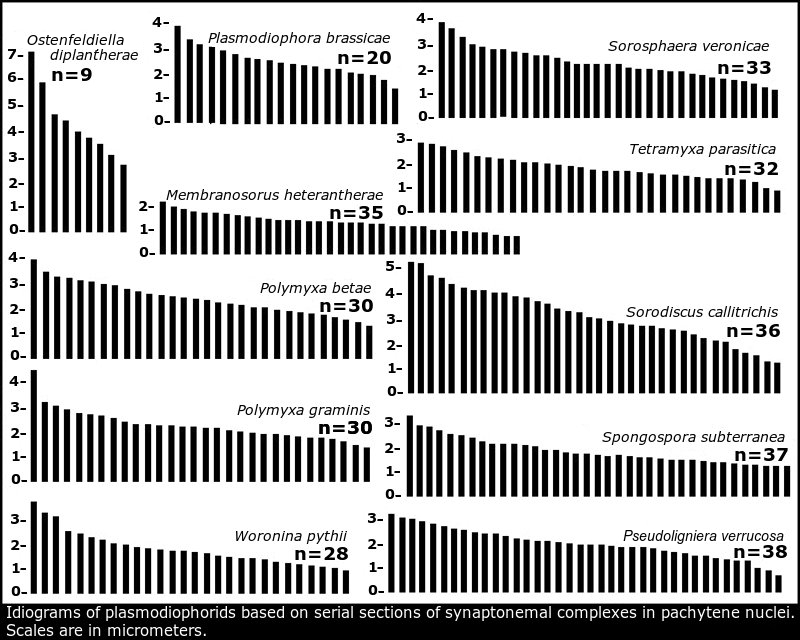
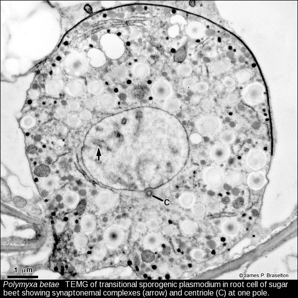
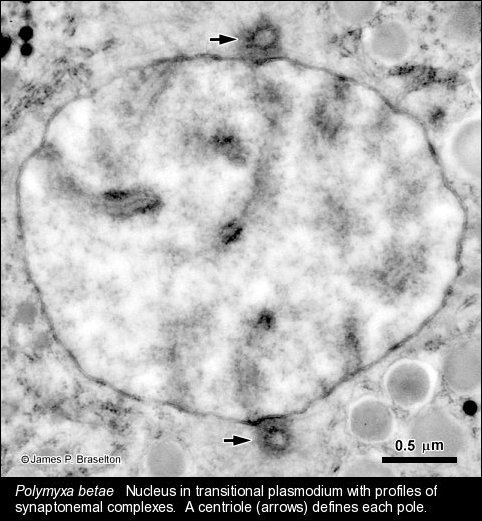
Barr (1979) pointed out that Ligniera and Polymyxa cannot be distinguished by their sporosori, but can, however, by their sporangial states. Karyotypic analysis revealed that the two species of Polymyxa have 30 synaptonemal complexes in pachynema whereas the former Ligniera verrucosa (now Pseudoligniera verrucosa) has 38.
Personal Comments
I thought that comparing the two species of Polymyxa with a Ligniera would be a good test of how well karyotyping with synaptonemal complexes could be used to characterize various plasmodiophorids. My opportunity to collect samples of P. betae came when I was visiting Stefan Buczacki at the then National Vegetable Research Station (NVRS) in Wellesbourne UK in 1982. He helped me make contact with Ian MacFarlane at Rothamsted Research Station in Harpenden, Hertfordshire UK.
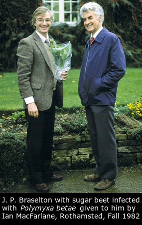
Anyone familiar with the plasmodiophorids would recognized that Ian MacFarlane had contributed to much of the fundamental information we had about the plasmos. For me it was an honor to visit him at Rothamsted and have him provide some of his sugar beet infected with P. betae. Ian was a very gracious host for the day and provided a wealth of information about his early work with the plasmodiophorids.
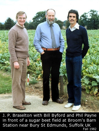
Ian and Stefan both introduced me to Bill Byford and Phil Payne at Broom's Barn near Bury St Edmunds in Suffolk. In addition to showing me the ins and outs of research into sugar beet, they treated me to lunch at a delightful country pub not far from the station. They also provided plants infected with P. betae.
The roots of sugar beet infected with P. betae were prepared for transmission electron microscopy at NVRS, but it was not until I returned to Ohio that I had time to serial section transitional sporogenic plasmodia to determine the karyotypes.
Once I returned to Ohio, Donald Barr, then at the Biosystematics Research Institute in Ottawa, Ontario, sent me a sample of dried roots of wheat that were infected with P. graminis. I used techniques for growing infected wheat on sand with nutrient solutions learned from Ian MacFarlane in his work with sugar beet.
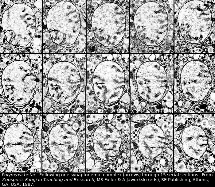
Both species of Polymyxa were beautiful in the prepared specimens. The synaptonemal complexes were better defined than those of Plasmodiophora brassicae, but the nuclei were larger and required more serial sections to completely make it through one nucleus. The material looked so good that I used 15 serial sections of a synaptonemal complex in P. betae for an illustration of the technique for karyotyping in Zoosporic Fungi in Teaching and Research, edited by M. S. Fuller and A. Jaworski, 1987.
Images of Polymyxa
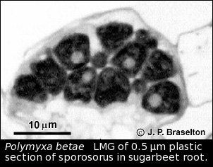
- Polymyxa resting spores, LMG
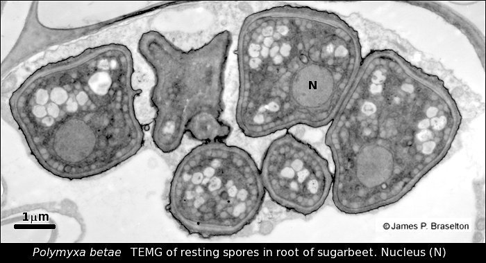
- Polymyxa betae resting spores, TEMG
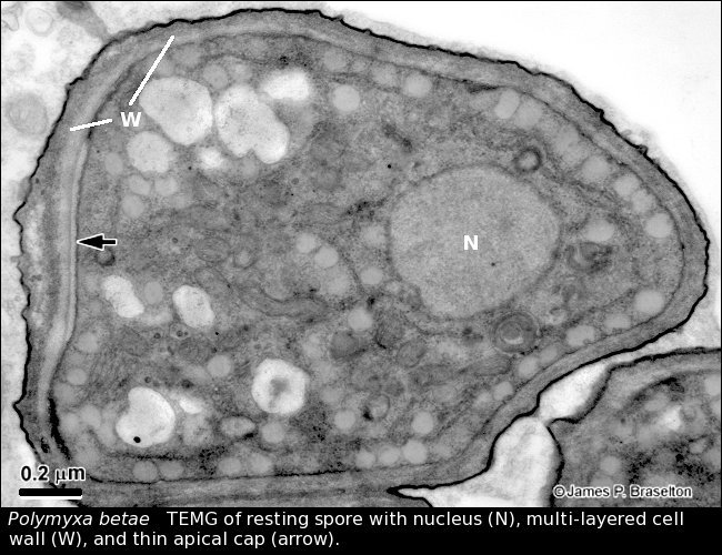
- Polymyxa betae resting spore, TEMG
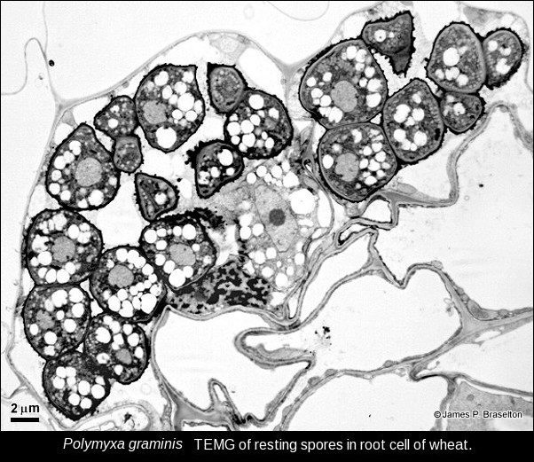
- Polymyxa graminis resting spores, TEMG
- Polymyxa sporogenic transitional plasmodium, TEMG
- Polymyxa transitional nucleus with synaptonemal complexs, TEMG
- Polymyxa serial sections of synaptonemal complex, TEMG
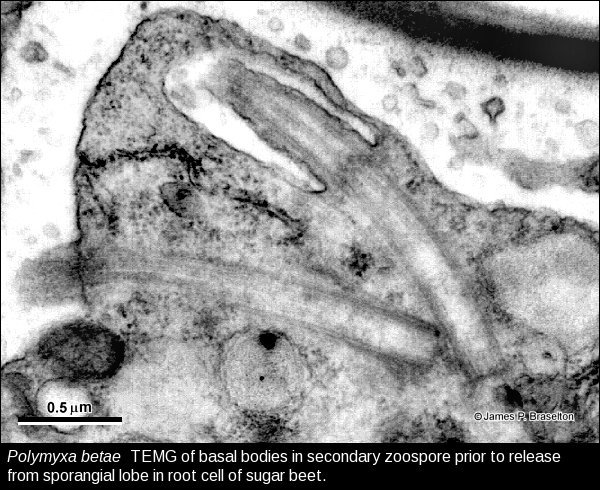
- Polymyxa zoospore, TEMG
Selected References for Polymyxa
- Barr, D. J. S. 1979. Morphology and host range of Polymyxa graminis, Polymyxa betae, and Ligniera pilorum from Ontario and some other areas. Can. J. Plant Path. 1: 85-94.
- _____ & P. M. E. Allan. 1982. Zoospore ultrastructure of Polymyxa graminis (Plasmodiophoromycetes). Can. J. Bot. 60: 2496-2504.
- Barr, K. J. & M. J. C. Asher. 1992. The host range of Polymyxa betae in Britain. Plant Pathology 41: 64-68.
- Braselton, J. P. 1983. Karyotypic analysis of Polymyxa betae (Plasmodiophoromycetes) based on serial thin sections of pachytene nuclei. Can. J. Bot. 61: 3202-3206.
- _____. 1984. Karyotypic analysis of Polymyxa graminis (Plasmodiophoromycetes) based on serial sections of synaptonemal complexes. Can. J. Bot. 62: 2414-2416.
- Ciafardini, G. & B. Marotta. 1989. Localization of fragile areas in walls of resting spores of Polymyxa betae. Can. J. Bot. 67: 3123-3126.
- D'Ambra, V. & R. Locci. 1971. Scanning electron microscopy investigations on Polymyxa betae Keskin. Riv. Pat. Veg. 7: 43-57.
- D'Ambra, V. & S. Mutto. 1975. Ultrastruttura di Polymyxa betae Keskin. Plasmodio, sporangio e cistorsoro. Riv. Pat. Veg. 11: 115-124.
- D'Ambra, V. & S. Mutto. 1977. The ultrastructure of Polymyxa betae zoospore exit-tube differentiation. Can. J. Bot. 55: 831-839.
- Desoignies, N., C. Stocco, C. Bragard, & A. Legrève. 2011. A new phenotype of Polymyxa betae in Arabidopsis thaliana. Eur. J. Plant Pathol. 131: 27-38.
- Falk, B. W. & J. E. Duffus. 1977. The first report of Polymyxa betae in the western hemisphere. Plant Dis. Reptr. 61: 492-494.
- Kanyuka, K., E. Ward, & M. J. Adams. 2003. Polymyxa graminis and the cereal viruses it transmits: a research challenge. Molecular Plant Pathology 4: 393-406.
- Keskin, B. 1964. Polymyxa betae n.sp., ein Parasit in den Wurzeln von Beta vulgaris Tournefort, besonders während der Jugendentwicklung der Zuckerrübe. Arch. Mikrobiol. 49: 348-374.
- Ledingham, G. A. 1939. Studies on Polymyxa graminis, n.gen. n.sp., a plasmodiophoraceous root parasite of wheat. Can. J. Res. C. 17: 38-51.
- Littlefield, L. J., P. Delfosse, J. H. Whallon, Z. M. Hassan, J. L. Sherwood, & D. V. R. Reddy. 1997. Anatomy of sporosori of Polymyxa graminis, the vector of Indian peanut clump virus, in roots of Sorghum bicolor. Can. J. Plant Pathol. 19: 281-288.
- Littlefield, L. J., J. H. Whallon, P. J. Doss, & Z. M. Hassan. 1998. Post infection development of Polymyxa graminis in roots of Triticum aestivum. Mycologia 90: 869-882.
- McGrann, G. R. D., M. K. Grimmer, E. S. Mutasa-Göttens, & M. Stevens. 2009. Progress towards the understanding and control of sugar beet rhizomania disease. Mol. Pl. Path. 10: 129-141.
- Tamada, T. & M. J. C. Asher. 2016. The Plasmodiophorid Protist Polymyxa betae. Pp. 135-153. In: Biancardi, E. & T. Tamada (eds.), Rhizomania, Springer International Publishing, Switzerland. Doi: 10.1007/978-3-319-30678-0_6