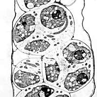
Plasmodiophorid Zoospores
Zoospores of plasmodiophorids have two whiplash flagella of unequal lengths. Primary zoospores are released from resting spores upon germination. Secondary zoospores are formed in thin-walled sporangia (a. k. a. zoosporangia).
Images of Plasmodiophorid Zoospores
- Spongospora zoospores, LMG
- Spongospora sporangial lobes with zoospores, TEMG
- Spongospora zoospore, TEMG
- Spongospora zoospore flagella, TEMG
- Polymyxa zoospore, TEMG
- Plasmodiophora sporangial lobes with zoospores, TEMG
- Diagram of zoospore
References for Plasmodiophorid Zoospores
- Aist, J. R. & P. H. Williams. 1971. The cytology and kinetics of cabbage root hair penetration by Plasmodiophora brassicae Wor. Can. J. Bot. 49: 2023-2034.
- Barr, D. J. S. & P. M. E. Allan. 1982. Zoospore ultrastructure of Polymyxa graminis (Plasmodiophoromycetes). Can. J. Bot. 60: 2496-2504.
- Buczacki, S. T. & C. M. Clay. 1984. Some observations on secondary zoospore development in Plasmodiophora brassicae. Trans. Br. Mycol. Soc. 82: 339-344.
- D'Ambra, V. & R. Locci. 1971. Scanning electron microscopy investigations on Polymyxa betae Keskin. Riv. Pat. Veg., Ser. IV, 7: 43-57.
- Kole, A. P. & A. J. Gielink. 1961. Electron microscope observations on the flagella of the zoosporangial zoospores of Plasmodiophora brassicae and Spongospora subterranea. Proc. Acad. Sci. Amsterdam Ser. C, 64: 157-161.
- Merz, U. 1992. Observations on swimming pattern and morphology of secondary zoospores of Spongospora subterranea. Plant Pathology 41: 490-494.
- Talley, M. R., C. E. Miller, & J. P. Braselton. 1978. Notes on the ultrastructure of zoospores of Sorosphaera veronicae. Mycologia 70: 1241-1247.