|
 |
|
Lawrence M. Witmer,
PhD
Professor of Anatomy
Chang Professor of Paleontology
Dept. of Biomedical Sciences
Heritage
College of Osteopathic Medicine
Life Science Building, Rm 123
Ohio University
Athens, Ohio 45701 USA
Phone: 740 593 9489
Fax: 740 593 2400
Email: witmerL@ohio.edu
|
|
|
|


 |
|
|
|
|
| |
 |
Rhinoceros Horn Growth &
Form
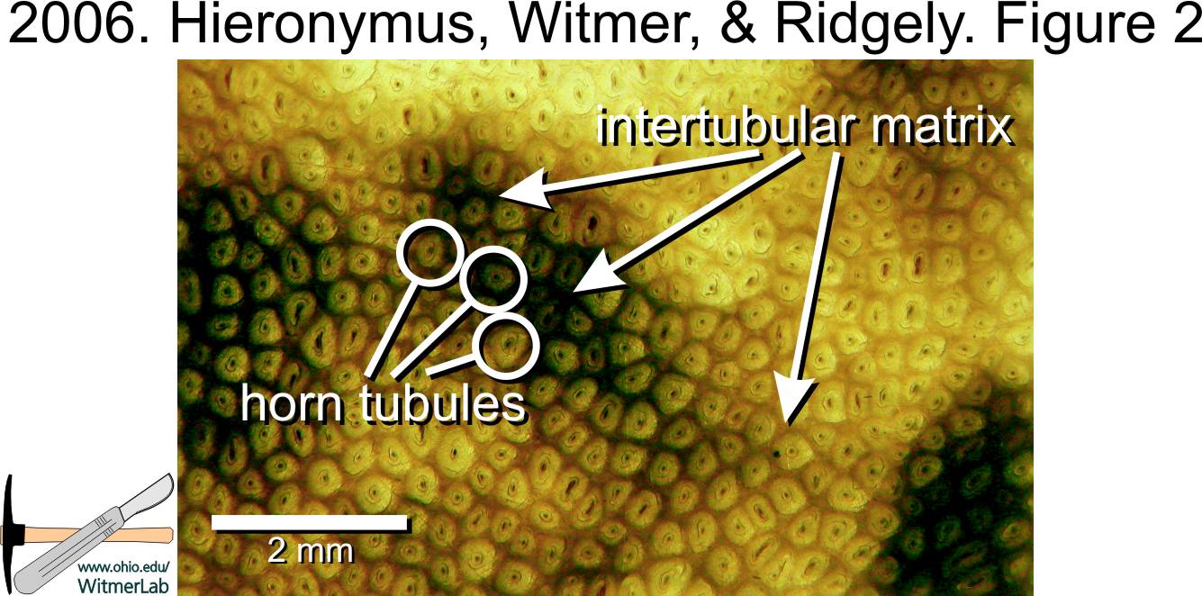
2006. Hieronymus, Witmer, and Ridgely. Journal of Morphology |
|
|
| |
|
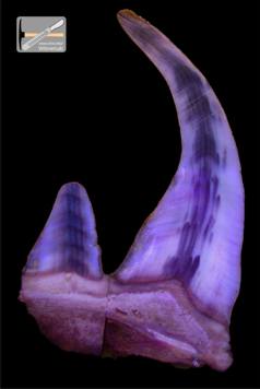
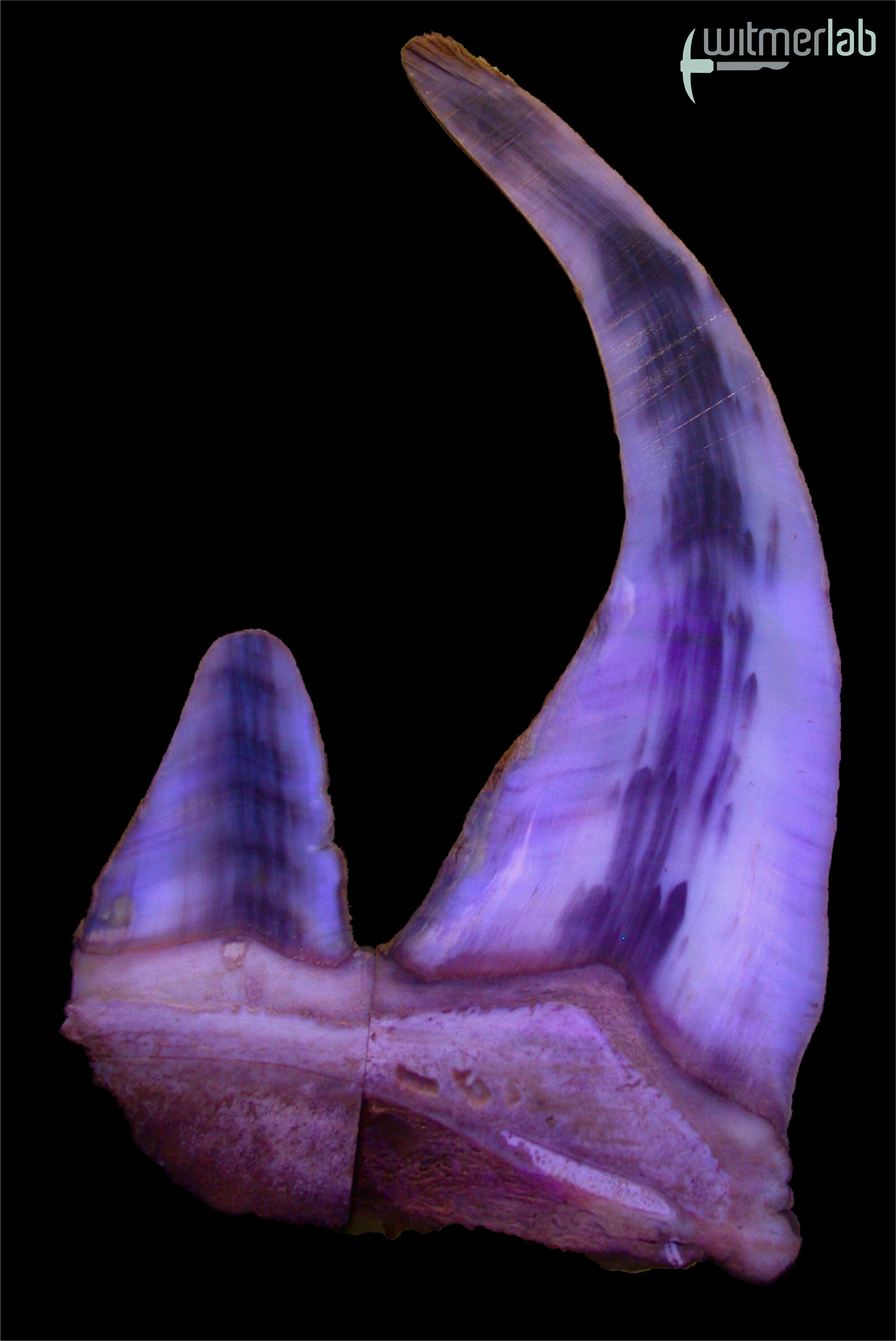
Click image to enlarge.
Image of sectioned rhino horn
photographed under ultraviolet light. |
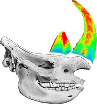
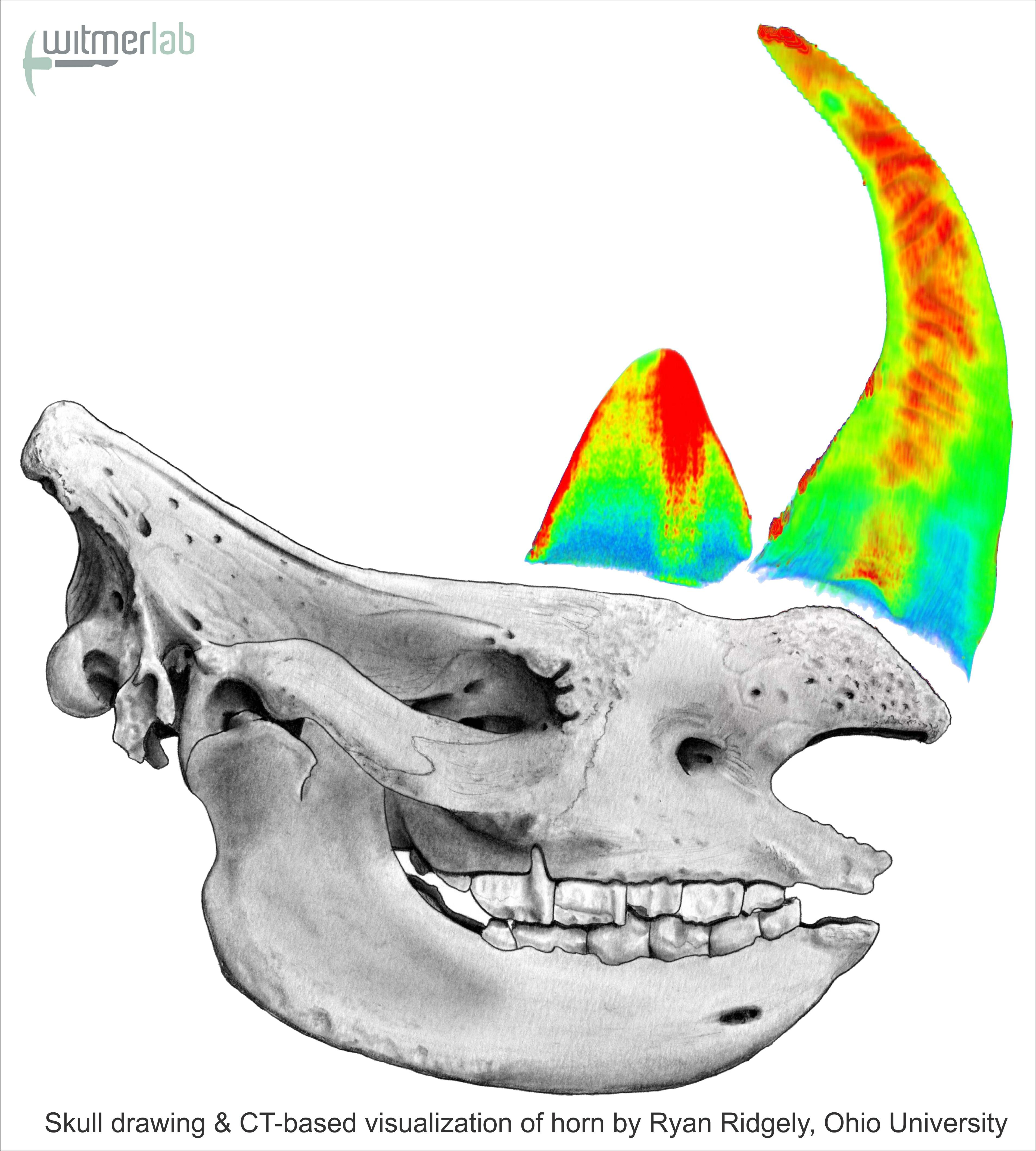
Click image to enlarge.
Drawing of rhino skull with CT-based
images of horns in place. Redder colors
represent denser portions. |
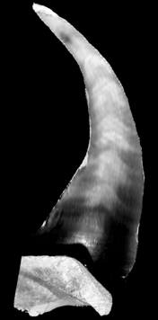
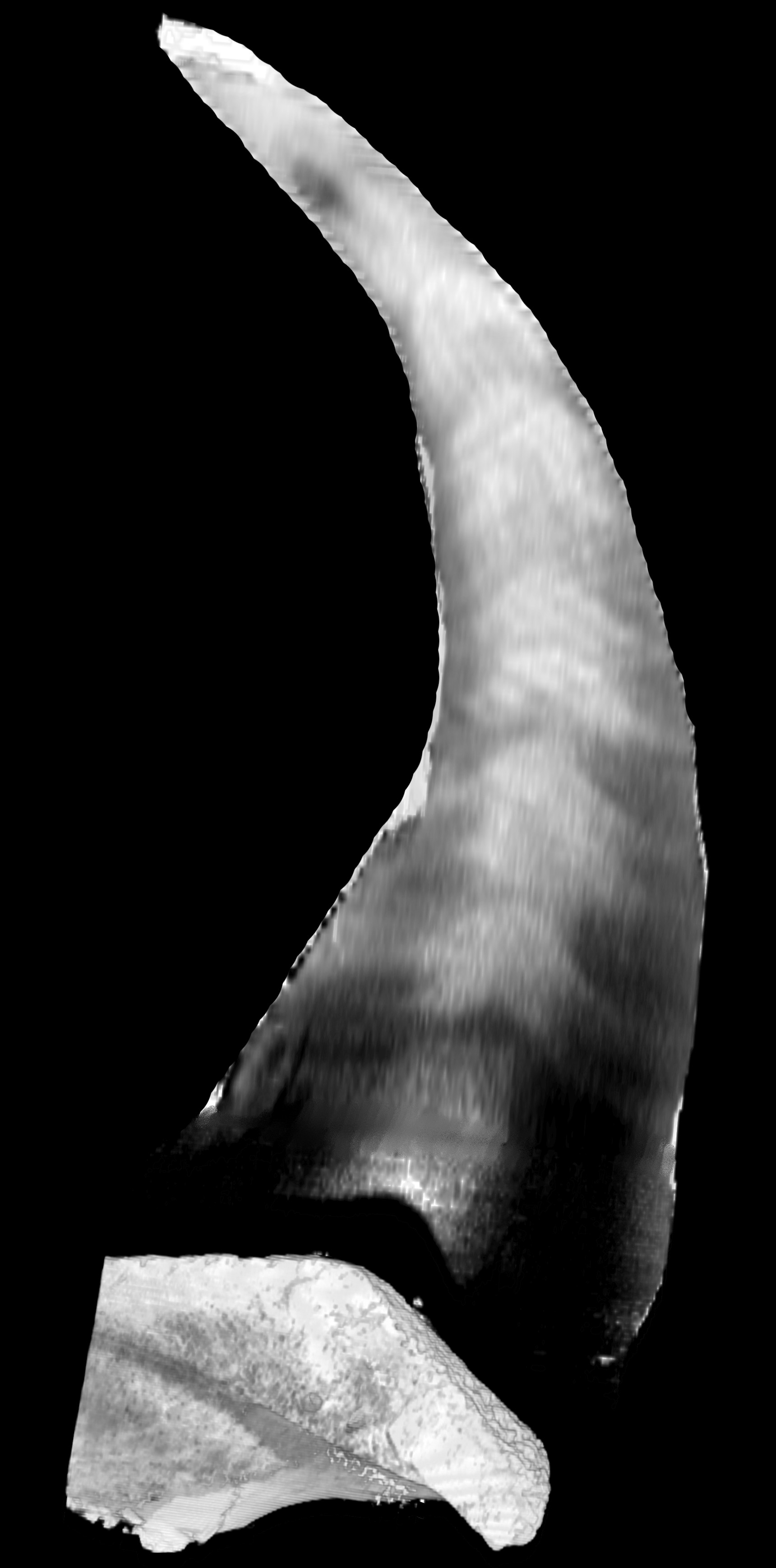
Click image to enlarge.
Image of CT slice through rhino horn.
Whiter areas represent denser portions. |
|
|
|
| |
Common Language Summary |
| |
CT
scanning sheds new light on the unique horns of rhinos.
Many kinds of projecting structures on animals are lumped
together under the term “horn.” There are horns on the heads of
cattle, on the beaks of hornbills, and on the faces of
chameleons, to name a few. In most cases, the external shape is
defined by a bony horn core, and the surface of the horn is only
a thin sheath of hardened keratins (the same materials in
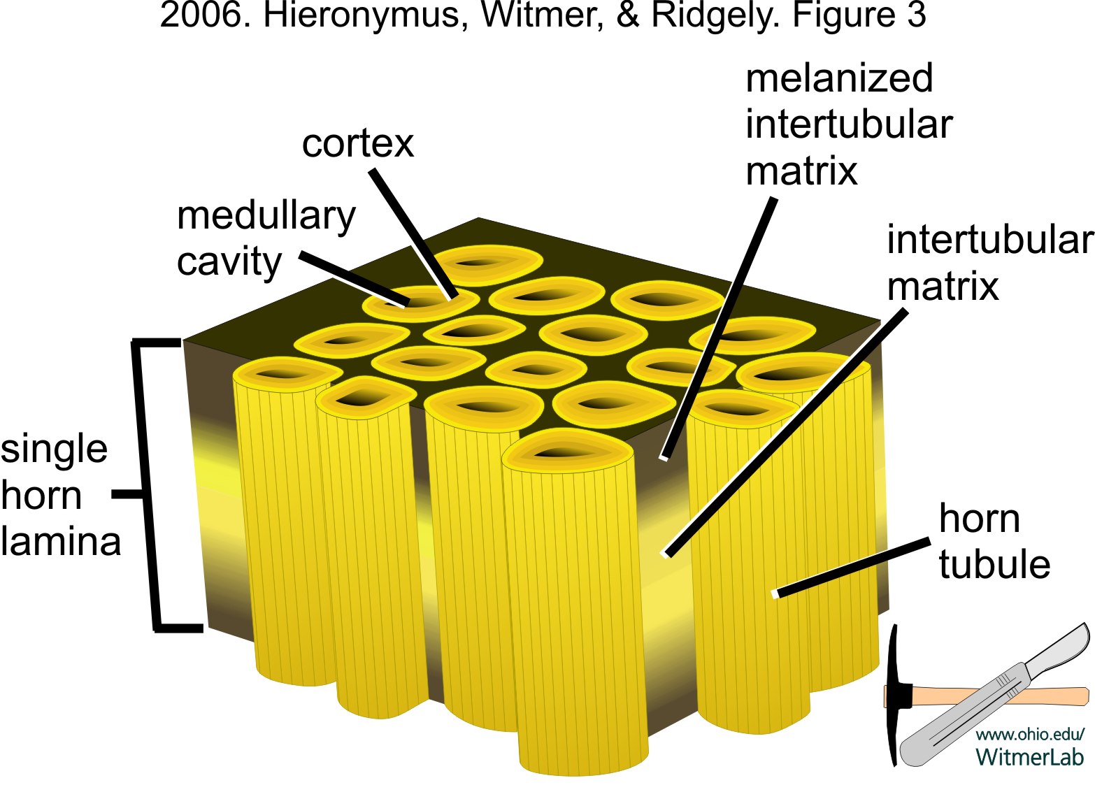
fingernails and hair). Rhinoceros are unique in having large
horns that are composed entirely of tiny keratin tubes embedded
in a keratinous matrix, without any bony core. CT scans and
cross-sections of rhino horns show a dense central region that
is reinforced by a combination of mineral (calcium) and melanin.
These two components make the center of the horn more resistant
to physical wear and breakdown due to UV light exposure whereas
the softer outer part is more easily worn away during use. Much
like a pencil, this difference in resistance leads to the
characteristic elongate and sharply pointed shape. Dark patches
of high mineral and melanin concentration within the horn match
up very well with measurements of annual horn growth in wild
rhinos, indicating that there are yearly “pulses” in the
deposition of these compounds. The tissue of rhinoceros horn
itself, with its tubules-within-matrix architecture, is very
similar to a number of other hardened skin appendages, including
the hooves of horses and cattle, whale baleen, and the tips of
bird beaks, all of which have evolved this skin architecture
independently. |
| |
|
| |
|
| |
note: Research
in the Witmer lab does not involve experimentation on live
animals. Specimens of modern animals used in research are
salvage specimens, obtained legally from commercial or
governmental sources. |
|