Karyogamy in Plasmodiophorids
Although meiosis has been extensively documented during cleavage of transitional sporogenic plasmodia into resting spores, location of karyogamy in plasmodiophorid life cycles remains a problem and merits further research. Several publications have interpreted some light and/or transmission electron microscopic images of nuclei close together as evidence for karyogamy, and articles by Buczacki & Moxham (1980) and Tommerup & Ingram (1971) suggested that karyogamy may occur in sporogenic plasmodia. Braselton (1995), however, questioned whether karyogamy was conclusively observed, which was consistent with Karling's (1968, page 28) summary of our knowledge of sexuality in plasmodiophorids: ". . . it is largely indirect and presumptive."
Kole's article (1954, page 14) summarized observations of fusion of zoospores, and pointed out that karyogamy was not observed in fused zoospores of Spongospora.
Personal Comments
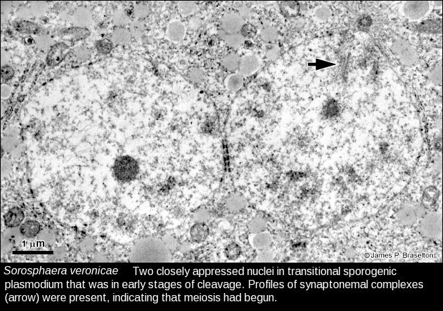
From the early 1970's through the beginning of 2000 (nearly 30 years!) I personally spent thousands of hours looking at thousands of sections of sporogenic plasmodia at light and transmission electron microscopic levels of Spongospora, Plasmodiophora, Sorosphaera, Sorodiscus, Tetramyxa, Woronina, Polymyxa, Membranosorus, and Ligniera, all the while hoping to see something that looked like karyogamy. Students under my direction and the direction of the late Charles E. Miller at Ohio University also were primed to look for karyogamy (discovery of it would have been their claim to fame). Obviously none of us ever saw anything that would confirm that karyogamy occurs in the plasmodiophorids. We came across transitional sporogenic plasmodia in Sorosphaera with nuclei that were appressed to each other. Upon closer observation, however, there were the beginnings of SCs in the nuclei and the nuclei were already the size of transitional nuclei with SCs; so all we could say was that the nuclei were close together, even lining up their nuclear pores. If fusion were occurring, we would have seen stages of karyogamy (e.g., channels between nuclei or interconnections of membranes) in that material, but we never did. When we found nuclei close together like shown in the image in the link above, we would go through the entire block of tissue to examine infections nearby to see if we could document karyogamy. Another brief moment when we thought solid evidence for karyogamy was found was when Dan Dylewski (Dylewski & Miller 1983) came across what he eventually determined to be stages of nuclear elimination during resting spore development in Woronina pythii.
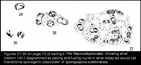
Earlier workers reports of karyogamy Figs. 27-30 (from Osborn 1911) on page 73 of Karling's 1968 The Plasmodioporales show what appear to be what we looked for all those years. If the drawings truly depict what Osborn observed, then that is close to documenting karyogamy. We can't be sure, however, until we see photographs showing similar images in material prepared by today's standards for microscopy.
There is still the possibility that karyogamy occurs in fused zoospores. There have been various reports over the years of tetraflagellate zoospores, suggesting that fusion of two biflagellate zoospores has occurred. As mentioned near the beginning of this page, however, Kole's article (1954, page 14) summarized observations of fusion of zoospores, and pointed out that karyogamy was not observed in fused zoospores of Spongospora.
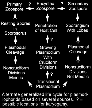
An alternate life cycle diagram that is based on several sources from the literature summarizes some of the possible locations for karyogamy in the plasmodiophorids.
References for Karyogamy