Home People Research Publications Teaching Synergy Gallery Join Us Contact Us

RESEARCH AND INSTRUMENT 


Funding: NIH-NHGRI-1R15HG009972

Super Resolution Imaging (Funded by National Human Genome Research Institute of NIH)


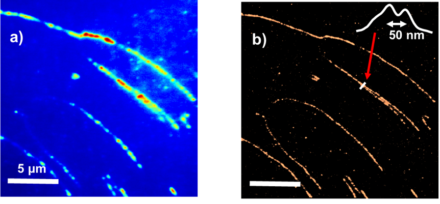
Super-resolution optical microscopy/nanoscopy has greatly extended our understanding of many systems, such as living cell structures and dynamics, and catalytically hot-spots on a nanoparticle. Super-resolution techniques allow the capture of images with a higher resolution than the diffraction limit, ~250 nm for fluorescence images with the highest resolution.

Due to the importance of these techniques to fundamental research, three pioneers in this field has been awarded the 2014 Nobel Prize in Chemistry. We are following these giants in applying the techniques in a various of area such as biophysics and materials science.

The group focuses on applying super-resolution imaging techniques to study molecular dynamics at interfaces. The PI has applied motion blur point accumulation for imaging in nanoscale topography (mbPAINT) to study DNA and protein imaging beyond the diffraction limit of light. The group continues to explore the opportunities in applying this technique in DNA super-resolution optical mapping.


Photophysics of Various Molecules/Nanomaterials


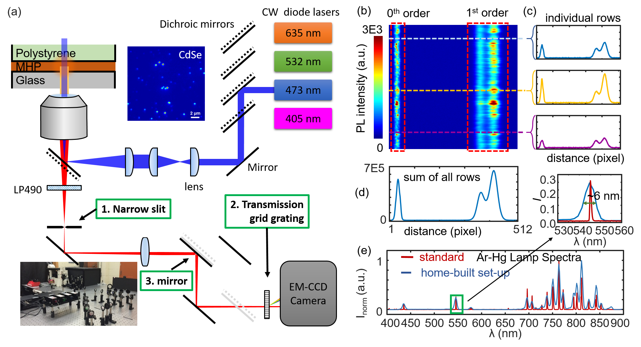
We are developing spectroscopic and microscopic solutions to study the photophysical properties of single molecules, single particles, and bulk thin films. For example, we have added FRET and hyperspectral microscopy functions to our super-resolution microscope and added gas environment and temperature control stages as well. We also collaborate with other groups in ultrafast spectroscopies of organic and inorganic dye systems. The above figure shows some examples of the setup, (a) left showing the scheme of the light path, (b, c, d) right showing measurement of perovskite thin film emmision, and (e) calibration of the spectromicroscopy.
 

Experiments and Simulations of Single-Molecule Kinetics


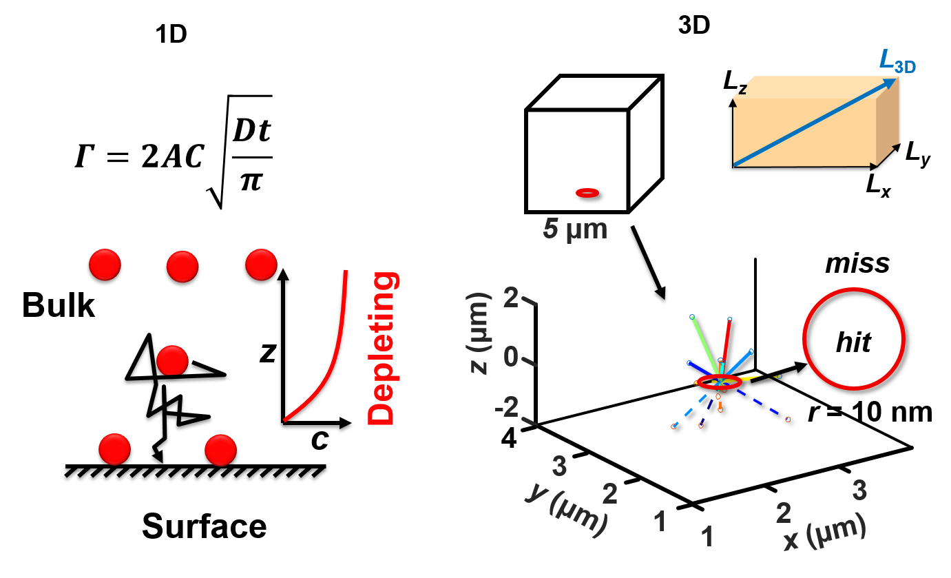
We also synthesize various nanoparticles and quantum dots for photoluminescence measurement and nanodevice fabrication. We use fluorescence microscopy to measure single-molecule diffusion/binding kinetics, single-molecule FRET kinetics, and single-particle fluorescence kinetics. And we use statistical methods to analyze these data. The left figure shows examples of using Monte Carlo simulations for the adsorption of molecules on a surface with 1D and 3D models. We are developing MATLAB codes for data analysis, fitting, and simulations on various kinetic systems, such as smFRET, reaction kinetics, and diffusion.


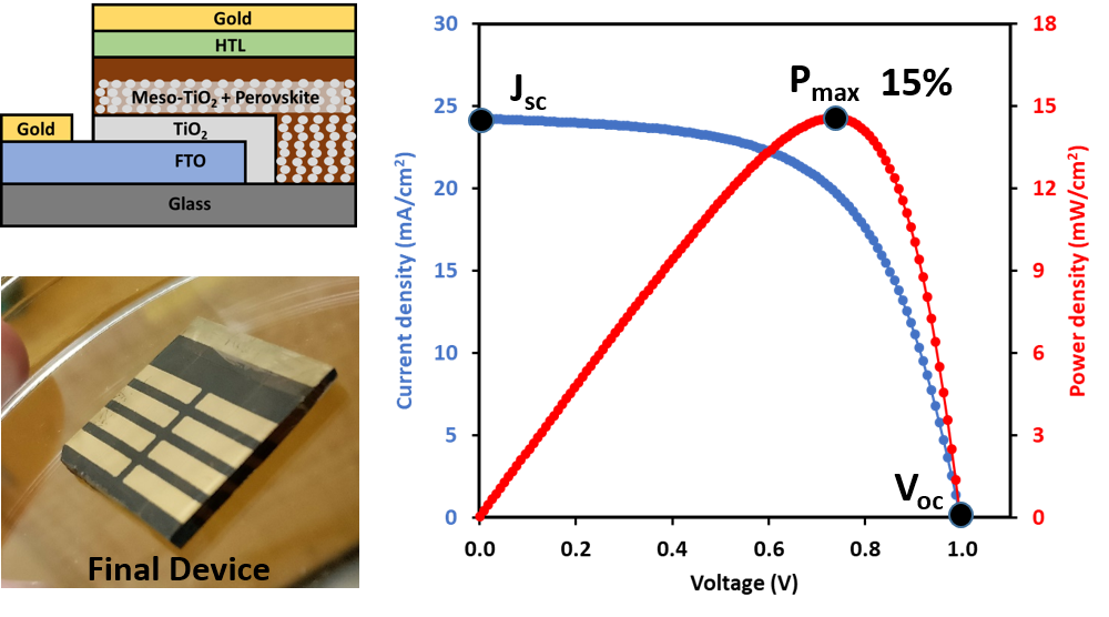
Solar Cells and nanofabrication


Our group is making thin-film solar cells and characterizing their interfacial chemical and electrochemical properties. The right figure shows a perovskite solar cell (PSC) fabricated and tested in our lab. PSC is a type of promising next generation solar cells.


Facilities and in-house instrumentations

Sharing space with the physical chemistry division, occupying about a third of the 4000 ft^2 lab space and 6 faculty and student offices in new chemistry building east wing of 3rd floor.

  • Wet lab : ~800 ft^2 equipted with benchtops, cabins, and three fume hoods, a laminar flow clean hood, water, sink, and an ultrapure water system.
  • Laser lab: ~400 ft^2 equipted with hang-on power rack, storage, and light control system.
  • PI and Student Offices across the hallway.
  • Have access to chemistry machine shop, chemistry stockroom, physics machine shop, and electronic shop.
  • Have access to NQPI and Ohio University shared equipment and facility.

In-house instruments:

  • A super-resolution spectro-microscope is ready for this project. More specifically, the microscope (Nikon TiU) is equipped with four lasers (405, 473, 532, and 635 nm),  a 1.49 NA, 100x, oil-immersion objective (Nikon CFI Apo), a 20x Nikon objective, filter sets, eyepieces, and a -100 oC cooled EMCCD detector (Andor iXon 897U). The working mode can be switched between total internal reflectance fluorescence (TIRF) and Epi-fluorescence wide-field mode.
  • A Raman/AFM/NSOM scanning microscope (AlphaSNOM, Witec GmbH) . This microscope incorporates confocal scanning Raman spectromicroscopy, atomic force microscopy (AFM),  and near-field scanning optical microscopy together in one microscope. The microscope has been further modified into a 4-π setup with two objectives focusing on the same plain. Several lasers have been connected to the microscope including CW lasers at 405 nm, 532 nm, and 980 nm, and a pulse laser at 532 nm. A high-resolution spectrometer (detector -100 oC) and a fast single-photon avalanche photodiode (APD) detector have been attached. A temperature-control microscope stage is equipped with this microscope. This microscope is obtained from Dr. Richardson, a recently retired colleague of the PI. This instrument is now shared in the physical chemistry division with Dr. Cimatu for teaching and research.
  • An atomic force microscope (MFP 3D AFM, Asylum Research shared with Dr. Cimatu for both teaching and research).
  • A differential scanning calorimeter (DSC shared with Dr. Cimatu for both teaching and research).
  • A fluorometer (Horiba shared with Dr. Cimatu for teaching and research)
  • MATLAB codes for data analysis of single-molecule and single-particle photoluminescence, MATLAB codes for ultrafast TA data global fitting, and PL lifetime fitting have been developed and tested in several publications.
  • A Dynamic light scattering spectrometer (DynaPro).
  • A VASE Ellipsometer (VASE HS-190, tunable wavelengths, also shared for teaching).
  • A single-photon counting spectrofluorometer (the best time resolution is 40 ps).
  • A gold sputter (Denton Vacuum) and a metal thermal evaporator.
  • A Plasma cleaner (Harrick Plasma).
  • Two spin coaters (MTI and Ossila).
  • A solar simulator (Abet).
  • A Keithley 2460 power source meter.
  • A Keithley 6514 electrometer.
  • A regular fluorescence microscope.
  • A UV-Vis spectrometer.
  • Other basic lab equipment such as balances, centrifuges, ovens, furnaces, refrigerators, safety chemical cabinets, sonicators, a probe sonicator, and a Thermo Barnstead E-Pure water purification system.

Home-built super-resolution fluorescence microscopy and spectro-microscopy


Witec Scanning Raman/SNOM/AFM spectroscopic and imaging system
Wavelength Scanning Fluorometer (can do lifetime measurement if we have an additional pulsed laser)
Tunable wavelength ellipsometer
Solar simulator
Cyclic voltameter and potential current meter
Laser cutter
Glovebox with a spin coater inside
Flow hood
Ultrapure water system, and Schlenk line (for orgnaic synthesis outside of glovebox) in the hood
Oven, centrifuge, balance, hoods, and etc.


Contact Us: Tel. No. : (740)-593-9768
e-mail: chenj (at) ohio (dot) edu