|
 |
|
Lawrence M. Witmer,
PhD
Professor of Anatomy
Chang Professor of Paleontology
Dept. of Biomedical Sciences
Heritage
College of Osteopathic Medicine
Life Science Building, Rm 123
Ohio University
Athens, Ohio 45701 USA
Email:
witmerL@ohio.edu
|
|
|
|


 |
|
|
|
| |
 |
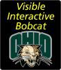
Visible
Interactive
Bobcat |
 |
|
Common
Language Summary
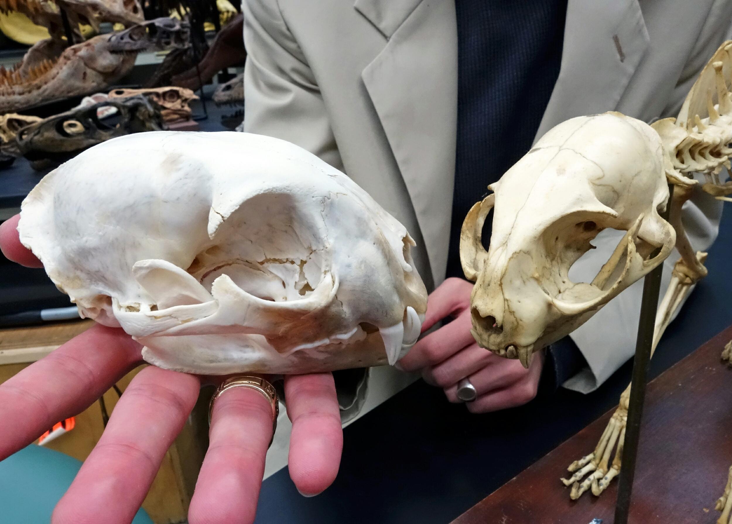
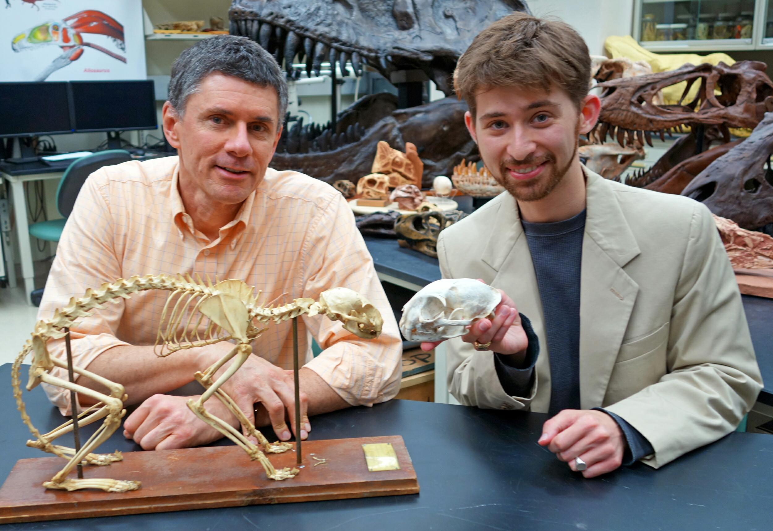
The Visible Interactive Bobcat.
This page presents our work on the 3D anatomical structure of
the head and skull of the bobcat, Lynx rufus. Bobcats are
among the few species of native wild cats in North America. The
OHIO Bobcats are the official sports mascot for Ohio
University, and although the species remains endangered in Ohio,
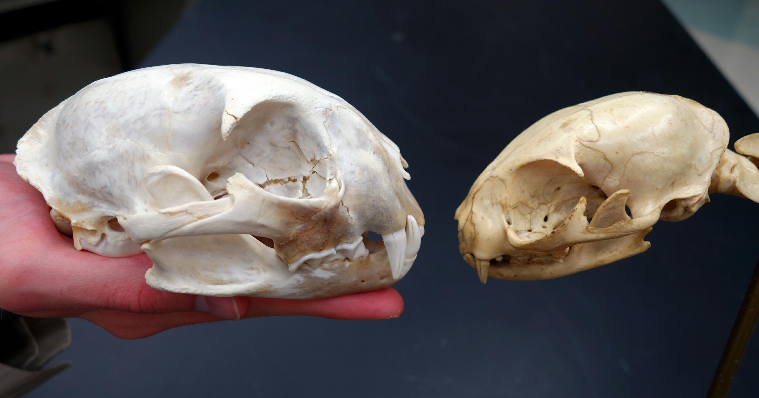
its numbers are recovering. The featured specimen here (OUVC
9576) is the skull of a large, adult bobcat from Athens County,
Ohio, supplied by the Ohio Department of Natural Resources. It
was a road kill victim, as evidenced by some skull fractures
visible in the imagery. The
resources on this page are outgrowths of our more technical work
and are intended to serve as STEM educational aids for K-12 and
undergraduate students, as well as for researchers. Work on this
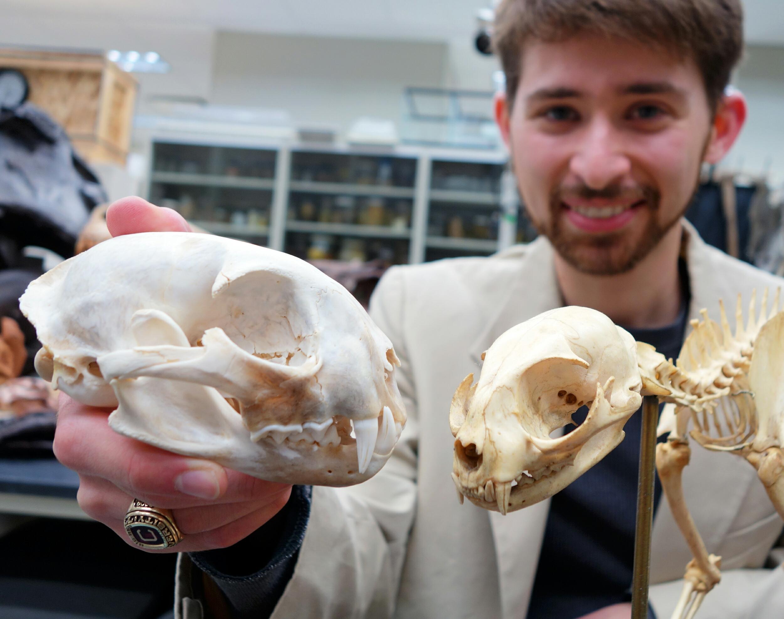
project was done by WitmerLab PhD student Donald Cerio. The skull
was µCT-scanned with a voxel size of 90 µm at the
OUµCT facility. Segmentation of anatomical structures was
done using Avizo; 3D PDFs were generated using Maya, Deep
Exploration, and Adobe Acrobat; and movies were made using
Avizo, QuickTime, and Adobe Premiere. GO BOBCATS!! |
|
|
|
|
 Download the µCT scan data
in DICOM format and 3D-printable STLs at MorphoSource.org
Download the µCT scan data
in DICOM format and 3D-printable STLs at MorphoSource.org
|
|
Sketchfab Animation |
Videos |
|
|
|
|
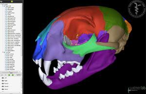
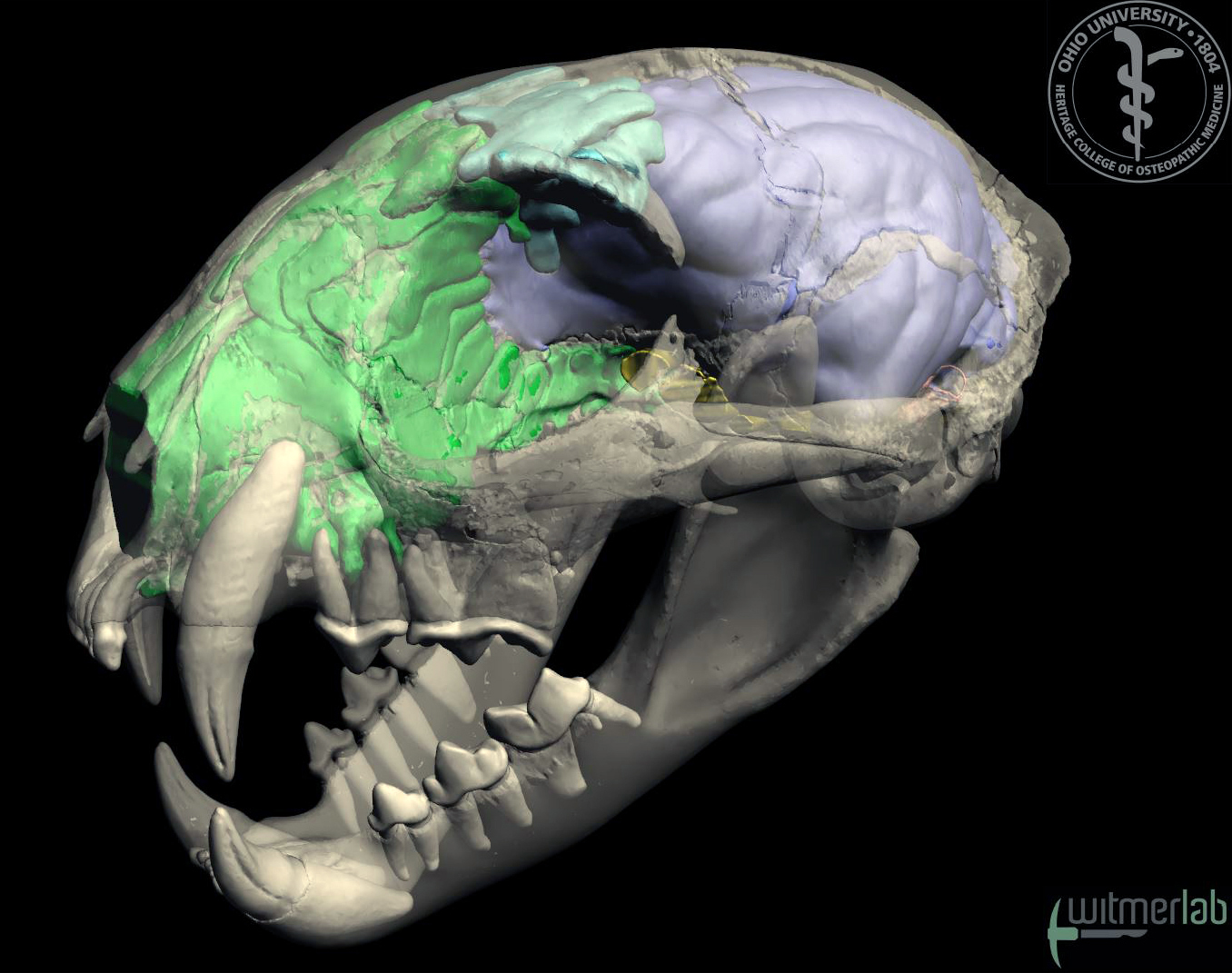
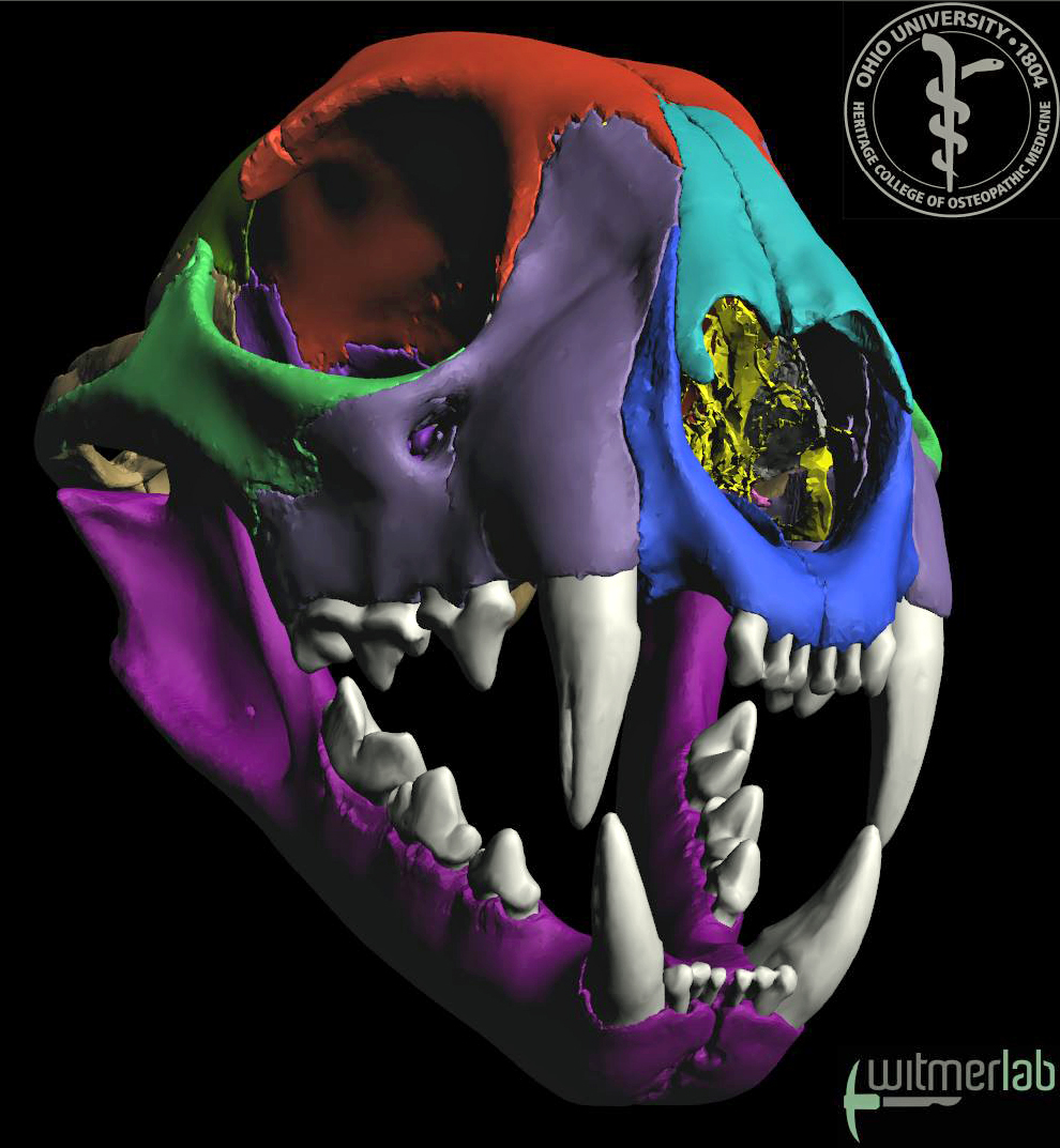
Labeled skull animation.
Animation of the skull of an
adult bobcat (Lynx rufus:
OUVC 9576), labeled to show the
individual bones of the skull.
Work on this project was done by
WitmerLab PhD student Donald
Cerio. The skull was µCT-scanned
with a voxel size of 90 µm at
the OUµCT facility. Segmentation
of anatomical structures was
done using Avizo; 3D PDFs were
generated using Maya, Deep
Exploration, and Adobe Acrobat;
and movies were made using
Avizo, QuickTime, and Adobe
Premiere.
•
Download a
46 MB QuickTime
version (HD: 1920x1080)
•
Download a
22 MB QuickTime
version (1280x720)
•
Download a
12 MB QuickTime
version (853x480)
•
Download an
8MB QuickTime
version (640x360) |
| |
| |
|
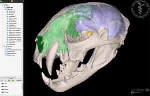
Labeled animation of skull, brain endocast, and inner ear. Animation of the skull of an adult bobcat (Lynx rufus: OUVC 9576), labeled to show the endocast of the brain cavity, labyrinth of the inner ear, paranasal air sinuses, and other soft tissues.. Work on this project was done by WitmerLab PhD student Donald Cerio. The skull was µCT-scanned with a voxel size of 90 µm at the OUµCT facility. Segmentation of anatomical structures was done using Avizo; 3D PDFs were generated using Maya, Deep Exploration, and Adobe Acrobat; and movies were made using Avizo, QuickTime, and Adobe Premiere.
• Download a 45 MB QuickTime version (HD: 1920x1080)
• Download a 23 MB QuickTime version (1280x720)
• Download a 12 MB QuickTime version (853x480)
• Download an 8 MB QuickTime version (640x360) |
|
|
|
|
|
|