Common
Language Summary
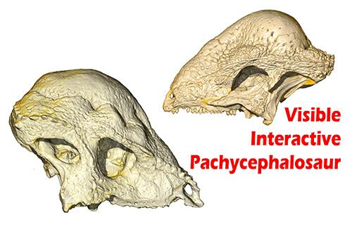
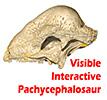
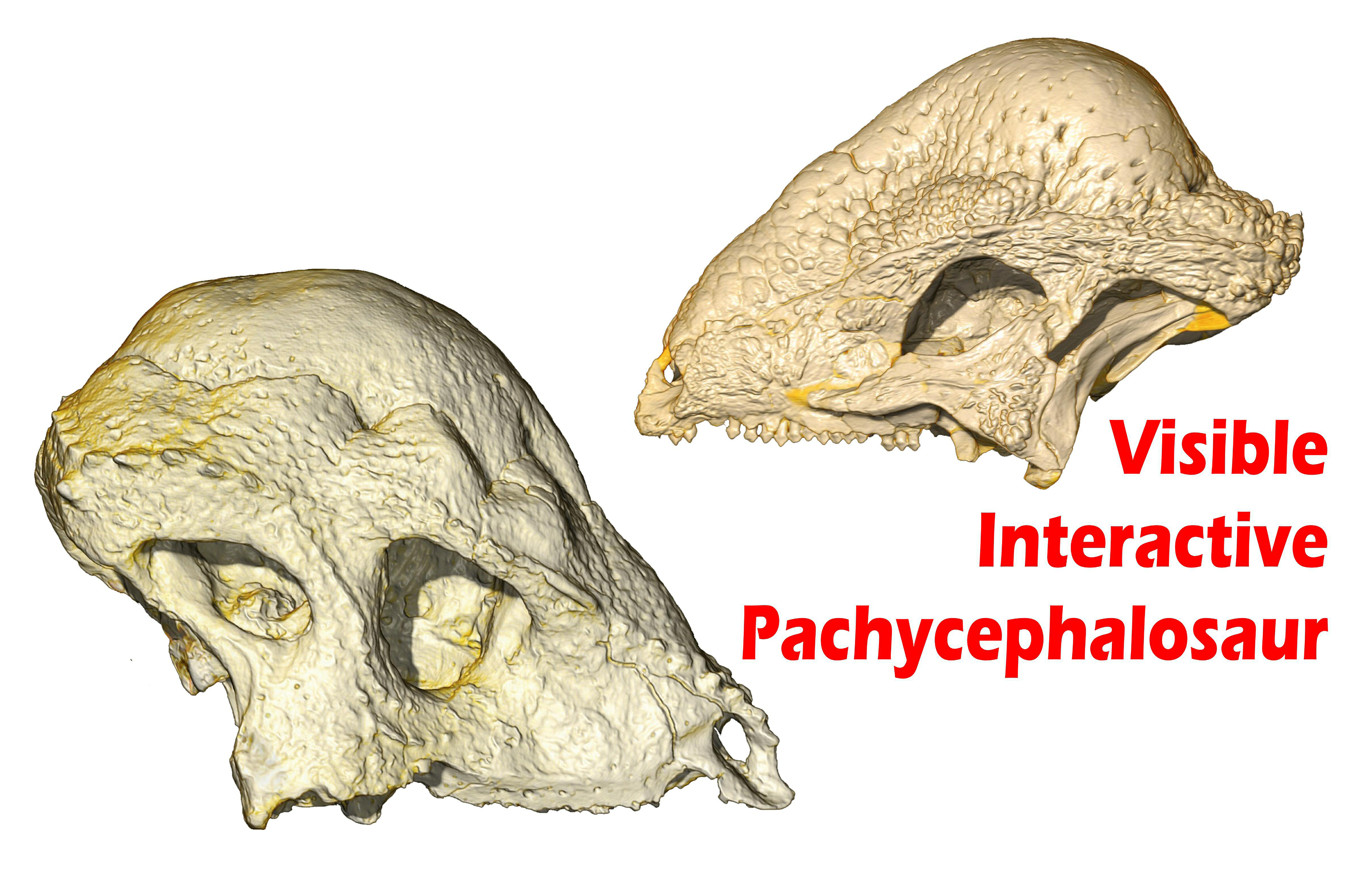
The Visible Interactive Pachycephalosaur.
This page presents our work on the 3D anatomical structure of
the skull of pachycephalosaurs, which are Cretaceous
ornithischian dinosaurs.
Three species are featured: (1) the holotype fossil skull of
Stegoceras validum (UALVP 2), collected from the Dinosaur
Park Formation of Alberta, Canada, and housed at the
University of Alberta;
(2) two fossil skulls of Sphaerotholus edmontonensis (MRF
360, MRF 361), collected from the Hell Creek Formation of
southwestern North Dakota and housed at the
Marmarth Research Foundation;
and (3) a cast of the holotype skull of Prenocephale prenes
(ZPAL MgD-1/104) collected from the Nemegt Formation of
Mongolia. These resources are outgrowths of our more technical
work and are intended to serve as STEM educational aids for K–12
and undergraduate students, as well as researchers. The
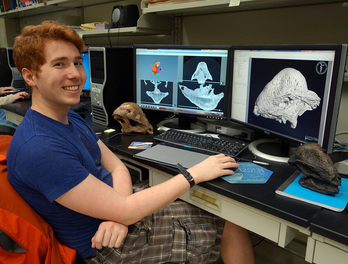
Stegoceras and Sphaerotholus specimens were CT
scanned at the University of Texas at Austin, and the cast of
Prenocephale was CT scanned at OhioHealth O’Bleness Hospital
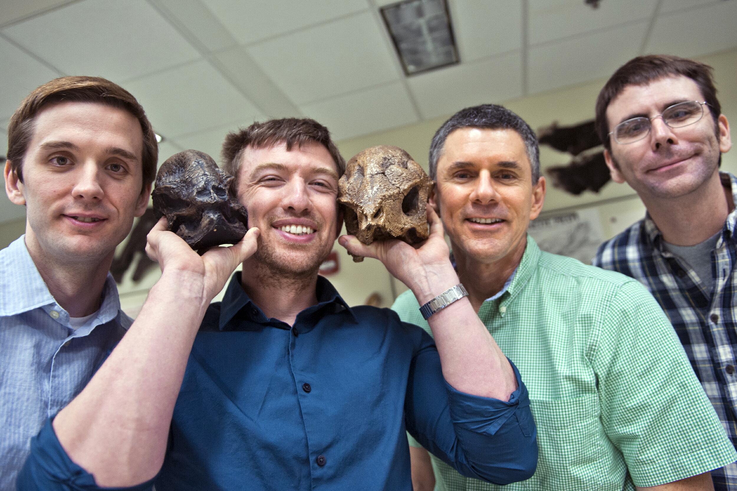
in Athens, Ohio. The scientific work on this project was done by
several WitmerLab members, including
Jason Bourke,
Ruger Porter, JP Nassif,
Ryan Ridgely, and
Larry Witmer.
More content will be added in the future. The project is funded
by grants from the National Science Foundation.
Check out
our other Visible Interactive Anatomy sites!
3D PDFs allow anyone with even the free Acrobat
Reader to interactively manipulate the 3D models that we
generate with powerful software like Avizo. The skull
and individual bones can be spun around, isolated, made
transparent, hidden, etc. The files can be saved to
your local computer. We provide each 3D PDF in different resolutions and files sizes to match your
interest and the power of your computer.
View our mini-tutorial. NOTE: Bugs in many browsers prevent them from running
3D PDFs in a browser window, so please save it to your
system and then launch it.
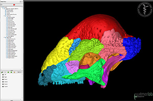
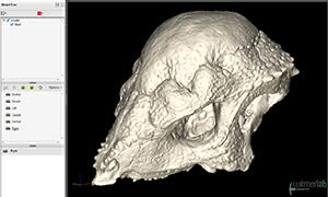
3D PDF of the skull of
the pachycephalosaurian dinosaur Stegoceras validum
(UALVP 2), collected from the Dinosaur Park Formation of
Alberta. The skull bones have all been digitally
isolated (and individually colored), allowing the skull
to be digitally dissected.
•
Download a
35 MB 3D PDF LARGE
•
Download a
17 MB 3D PDF MEDIUM
•
Download a
9 MB 3D PDF SMALL
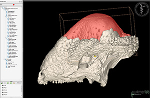
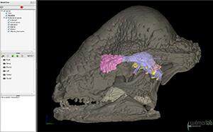
3D PDF of the skull of
the pachycephalosaurian dinosaur Stegoceras validum
(UALVP 2), collected from the Dinosaur Park Formation of
Alberta. The skull bones have all been digitally
isolated so that the skull can be digitally
dissected. The olfactory turbinates, brain
endocast, endosseous labyrinth, and neurovasculature
•
Download a
48 MB 3D PDF LARGE
•
Download a
24 MB 3D PDF MEDIUM
•
Download a
12 MB 3D PDF SMALL
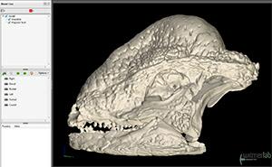
3D PDF of the skull of
the pachycephalosaurian dinosaur Stegoceras validum
(UALVP 2), collected from the Dinosaur Park Formation of
Alberta. Note: the skull has been digitally prepared to
be matrix-free, so use the slice tools in Acrobat to
view the internal anatomy.
•
Download a 28 MB 3D PDF
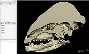
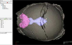
3D PDF of the skull of
the pachycephalosaurian dinosaur Stegoceras validum
(UALVP 2), collected from the Dinosaur Park Formation of
Alberta, showing the olfactory turbinates, brain
endocast, endosseous labyrinth, and neurovasculature.
•
Download a
32 MB 3D PDF
3D PDF of the skull of the pachycephalosaurian dinosaur
Sphaerotholus edmontonensis
(MRF 360), collected from the Hell Creek Formation of
North Dakota, showing the olfactory turbinates, brain
endocast, endosseous labyrinth, and neurovasculature.
•
Download a
29 MB 3D PDF
3D PDF of a cast of the skull of
the pachycephalosaurian dinosaur Prenocephale prenes
(ZPAL MgD-1/104), the holotype specimen collected from
the Upper Cretaceous Nemegt Formation of Mongolia.
•
Download a
15 MB 3D PDF
Photos
Animation of sagittal microCT
slices through the skull of the
pachycephalosaurian dinosaur
Stegoceras validum (UALVP
2). The specimen was scanned at
the University of Texas
High-Resolution CT Facility (UTCT)
in March 2010.
•
Download a
23 MB QuickTime
version (HD: 1920x1080)
Animation of horizontal microCT slices through the skull of the pachycephalosaurian dinosaur Stegoceras validum (UALVP 2). The specimen was scanned at the University of Texas High-Resolution CT Facility (UTCT) in March 2010
• Download a 15 MB QuickTime version (HD: 1920x1080)
Animation of axial microCT slices through the holotype skull of the pachycephalosaurian dinosaur Stegoceras validum (UALVP 2). The specimen was scanned at the University of Texas High-Resolution CT Facility (UTCT) in March 2010.
• Download a 15 MB QuickTime version (HD: 1920x1080)
Animation of sagittal microCT slices through the skull of the pachycephalosaurian dinosaur Sphaerotholus edmontonensis (MRF 360). The specimen was scanned at the University of Texas High-Resolution CT Facility (UTCT) in July 2009.
• Download a 37 MB QuickTime version (HD: 1920x1080)
Animation of horizontal microCT slices through the skull of the pachycephalosaurian dinosaur Sphaerotholus edmontonensis (MRF 360). The specimen was scanned at the University of Texas High-Resolution CT Facility (UTCT) in July 2009.
• Download a 27 MB QuickTime version (HD: 1920x1080)
Animation of axial microCT slices through the skull of the pachycephalosaurian dinosaur Sphaerotholus edmontonensis (MRF 360). The specimen was scanned at the University of Texas High-Resolution CT Facility (UTCT) in July 2009.
• Download a 19 MB QuickTime version (HD: 1920x1080)
Witmer is responsible for
the content of the website. Content provided here is for
educational and research purposes only, and may not be used for
any commercial purpose without the permission of
L. M. Witmer and other
relevant parties.