Common
Language Summary
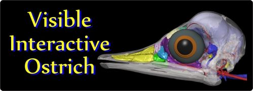
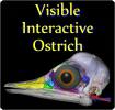
The Visible Interactive Ostrich.
This page presents our work on the 3D anatomical
structure of the head and skull of the ostrich,
Struthio camelus. These resources are
outgrowths of our more technical work and are intended to serve
as STEM educational aids for K-12 and undergraduate students, as
well as for researchers. WitmerLab has been working on ostrich
cephalic anatomy for many years, and work on this site draws
from the efforts of many lab members, most recently Ruger
Porter, Cheyenne Romick,
Ashley Morhardt, and
Jason Bourke.
CT scanning was done at both the
OUµCT facility and
OhioHealth
O'Bleness Hospital. Segmentation of anatomical structures was
done using Amira and Avizo, 3D PDFs were generated using Deep
Exploration and Adobe Acrobat, and movies were made using
QuickTime or Adobe Premiere.
Featured specimens
here include a two-month-old ostrich (OUVC 10519) that has been
the focus of more recent efforts as well as an adult specimen
(OUVC 10491) that was published in a
2008 Witmer & Ridgely article in the Anatomical Record.
Check out
our other Visible Interactive Anatomy sites!
3D PDFs
Videos
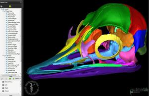
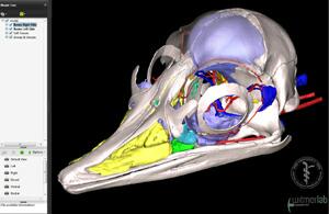
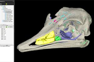
3D PDFs allow anyone with even the free Acrobat
Reader to interactively manipulate the 3D models that we
generate with powerful software like Avizo. The skull
and individual bones can be spun around, isolated, made
transparent, hidden, etc. The files can even be saved to
your local computer. We provide each 3D PDF in different resolutions and files sizes to match your
interest and the power of your computer.
View our mini-tutorial. NOTE: Bugs in many browsers prevent them from running
3D PDFs in a browser window, so please save it to your
system and then launch it.
Labeled animation of head
anatomy. Animation of the
head and skull anatomy of a
two-month-old ostrich (Struthio
camelus, OUVC 10519),
labeled to show the individual
bones of the skull, as well as
the anatomy of the airway,
paranasal air sinuses, brain
endocast, eyeball, cephalic
vasculature, and endosseous
labyrinth of the inner ear. The
arteries and veins of the head
were injected with a
barium/latex medium, and then
the head was microCT scanned at
45 µm and 90 µm voxel
resolutions. The 3D
visualization work was done in
Avizo, CorelDraw, and Adobe
Premiere by William Porter,
Cheyenne Romick, Ashley
Morhardt, and Jason Bourke.
•
Download a 43 MB QuickTime
version (HD: 1920x1080)
•
Download a 25 MB QuickTime
version (1280x720)
•
Download a 20 MB QuickTime
version (853x480)
•
Download a 11 MB QuickTime
version (640x360)
Axial CT slices. Older movie made in 2008 depicting CT scan slices in the axial (transverse) plane of the head of Struthio camelus (OUVC 10491). The head was scanned at O'Bleness Memorial Hospital on a GE LightSpeed Ultra Multislice CT scanner. Rendered using Amira and QuickTime by Ryan Ridgely. This movie derives from the 2008 Witmer & Ridgely article in the Anatomical Record..
• Download a 4.2 MB QuickTime version (640x568)
Sagittal CT slices. Older movie made in 2008 depicting CT scan slices in the sagittal plane of the head of Struthio camelus (OUVC 10491). The head was scanned at O'Bleness Memorial Hospital on a GE LightSpeed Ultra Multislice CT scanner. Rendered using Amira and QuickTime by Ryan Ridgely. This movie derives from the 2008 Witmer & Ridgely article in the Anatomical Record..
• Download a 4.0 MB QuickTime version (944x460)
Horizontal CT slices. Older movie made in 2008 depicting CT scan slices in the horizontal plane of the head of Struthio camelus (OUVC 10491). The head was scanned at O'Bleness Memorial Hospital on a GE LightSpeed Ultra Multislice CT scanner. Rendered using Amira and QuickTime by Ryan Ridgely. This movie derives from the 2008 Witmer & Ridgely article in the Anatomical Record..
• Download a 4.0 MB QuickTime version (950x462)
This website provides supplementary information as an
adjunct to published paper. Witmer, with the skilled
assistance of
Ryan Ridgely, is responsible for
the content of the website. Content provided here is for
educational and research purposes only, and may not be used for
any commercial purpose without the permission of
L. M. Witmer and other
relevant parties.