Common
Language Summary
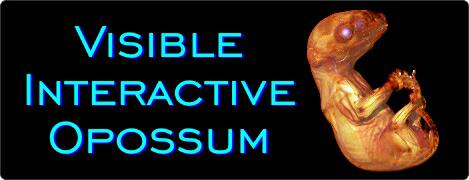
The Visible
Interactive Opossum. This page presents our work on the 3D
anatomical structure of the Virginia opossum, Didelphis
virginiana. Since we’re not currently doing real research on
opossum anatomy—we just used these long-deceased specimens in
experiments to hone a technique—we’re giving all the data
away…free and open-access! These resources are outgrowths of
our more technical work and are intended to serve as STEM
educational aids for K-12 and undergraduate students, but we
provide the full µCT-scan datasets for use by researchers. Our
featured specimens are two approximately 21-day-old opossum
pouch young (OUVC 10713 and OUVC 10716). Formalin-fixed
specimens were soaked in 20% sucrose solution for 24 hours and
then were submerged in 1.25% iodine Lugol’s solution for 48 hours. They
were then scanned on the OUµCT scanner at a resolution of 45 µm
(0.045 mm). Slice movies were produced in Avizo 7.1. Movies were
edited in QuickTime 7.7.3 and Adobe Premiere Pro CS6. The work
on this project was done by
Ashley Morhardt as part of her doctoral dissertation in the
WitmerLab. Ashley developed these resources as part of an
ongoing effort to visualize soft-tissue anatomy in CT data using
a pre-scan bath of Lugol’s iodine solution (iodine potassium
iodide — I2KI). During pre-scan baths, specimens are soaked in
iodine solution, allowing iodine (radiopaque halogen) atoms to diffuse
from the solution and into a specimen’s soft tissues, making
them visible on scan data. Content for this site includes DICOM
data for both specimens, slice movies for OUVC 10713, and a 3D
PDF of a skeleton scanned prior to immersion in Lugol’s iodine.
Check out
our other Visible Interactive Anatomy sites!
FREE, OPEN-ACCESS
µCT SCAN DATA
Download the raw
DICOM scan data for two specimens of the same age.
Note these are large ZIP files that
will expand to 400-500 MB:
OUVC 10713 (256 MB)
OUVC 10716 (295 MB)
3D PDFs
Videos
3D PDFs allow anyone with even the free Acrobat
Reader to interactively manipulate the 3D models that we
generate with powerful software like Avizo. The skeleton can be spun around, made
transparent, hidden, etc. The files can even be saved to
your local computer. We provide the 3D PDF in different resolutions and files sizes to match your
interest and the power of your computer.
View our mini-tutorial. NOTE: Bugs in many browsers prevent them from running
3D PDFs in a browser window, so please save it to your
system and then launch it.
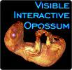
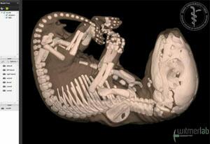
Slice
animation of head
anatomy. Animation of
microCT slices through a pouch
young of a Virginia opossum (Didelphis
virginiana, OUVC 10713).
This specimen is a ~21-day-old joey that was fixed in formalin,
immersed in a solution of iodine
potassium iodide (25% Lugol's)
to provide soft-tissue
resolution, and then microCT-scanned
at 45 µm. The animation runs
through slices in all three
planes, first through the
horizontal plane, followed by
transverse and then sagittal
slices. The 3D visualization
work was done in Avizo,
QuickTime, and Adobe Premiere by
Ashley Morhardt. Note: this
movie basically includes the
content of the other three slice
movies, putting it all together
in one place.
•
Download a
65 MB QuickTime
version (HD: 1920x1080)
•
Download a
32 MB QuickTime
version (1280x720)
•
Download a
17 MB QuickTime
version (853x480)
•
Download an 11 MB QuickTime
version (640x360)
Witmer is responsible for
the content of the website. Content provided here is for
educational and research purposes only, and may not be used for
any commercial purpose without the permission of
L. M. Witmer and other
relevant parties.