|
 |
|
Lawrence M. Witmer,
PhD
Professor of Anatomy
Chang Professor of Paleontology
Dept. of Biomedical Sciences
Heritage
College of Osteopathic Medicine
Life Science Building, Rm 123
Ohio University
Athens, Ohio 45701 USA
Email:
witmerL@ohio.edu
|
|
|
|


 |
|
|
|
|
|
Common
Language Summary
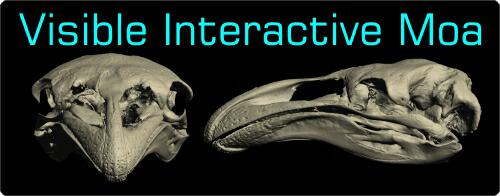
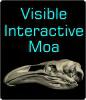
The Visible Interactive Moa.
This page presents our work on the 3D anatomical
structure of the head and skull of the extinct South Island
Giant Moa, Dinornis robustus. These resources are
outgrowths of our more technical work and are intended to serve
as STEM educational aids for K–12 and undergraduate students, as
well as researchers. Moa were a group of giant flightless birds
endemic to New Zealand until the 1400s, when they went extinct.
Members of the Dinornis robustus species were the largest
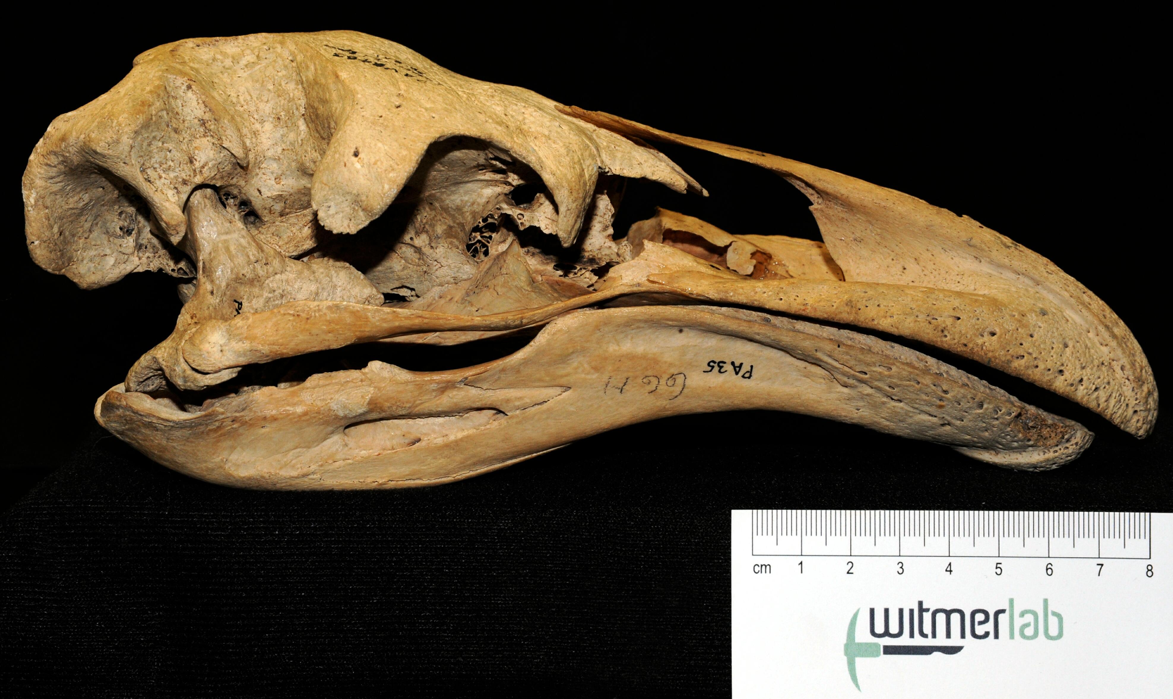
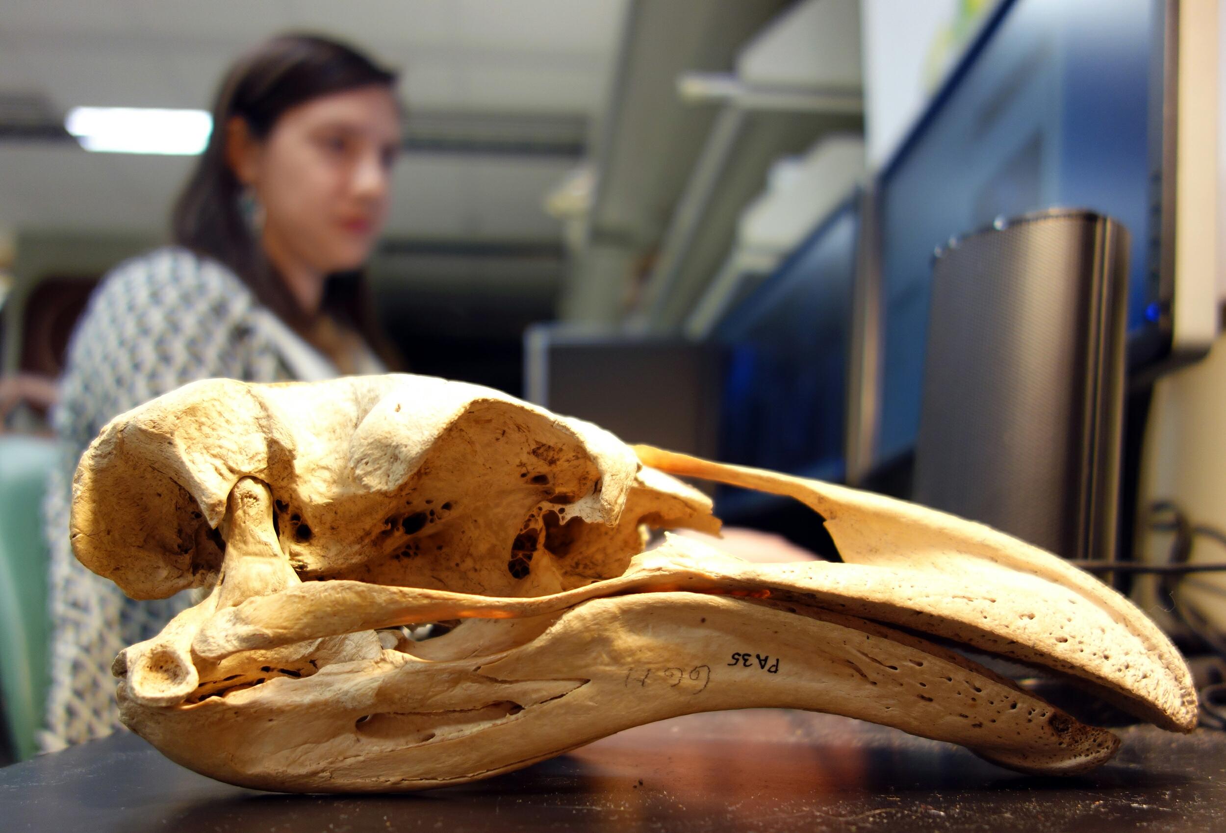
moa discovered to date. The featured specimen, FMNH PA 35,
belongs to the Field Museum of Natural History in Chicago and is
a mature female. It was collected in 1949 in the Pyramid Valley
swamp near Christchurch, New Zealand, for the Canterbury Museum (thanks
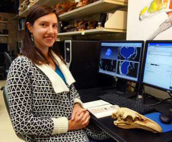
go to Paul Scofield for information on this specimen). Work on
this project was primarily done by WitmerLab PhD student
Catherine Early, with WitmerLab PhD candidate Ruger Porter
contributing details of the vasculature. The bones of the
braincase, mandible, and face were CT scanned at
OhioHealth O’Bleness Hospital,
Athens, Ohio,
at a slice thickness of 300 µm, and the quadrate bone was
scanned at the OUµCT
facility at a slice thickness of 45 µm. Segmentation of
anatomical structures was done using Avizo, 3D modeling was done
using Maya, 3D PDFs were generated using Deep Exploration and
Adobe Acrobat, and movies were made using QuickTime and Adobe
Premiere. |
|
|
|
|
|
3D PDFs |
Videos |
3D PDFs allow anyone with even the free Acrobat
Reader to interactively manipulate the 3D models that we
generate with powerful software like Avizo. The skull
and individual bones can be spun around, isolated, made
transparent, hidden, etc. The files can even be saved to
your local computer. We provide each 3D PDF in different resolutions and files sizes to match your
interest and the power of your computer.
View our mini-tutorial.
NOTE: Bugs in many browsers prevent them from running
3D PDFs in a browser window, so please save it to your
system and then launch it.
|
|
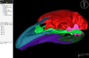 |
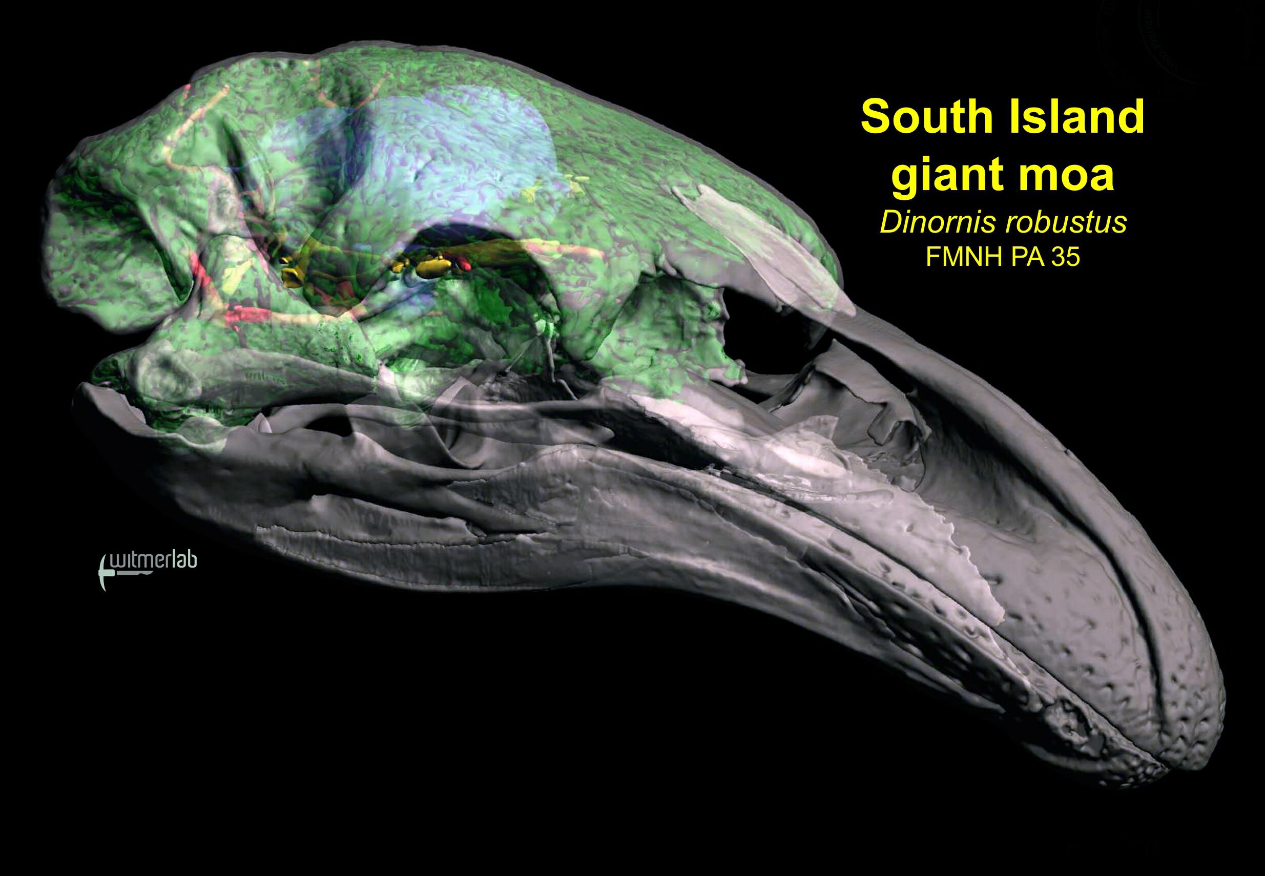
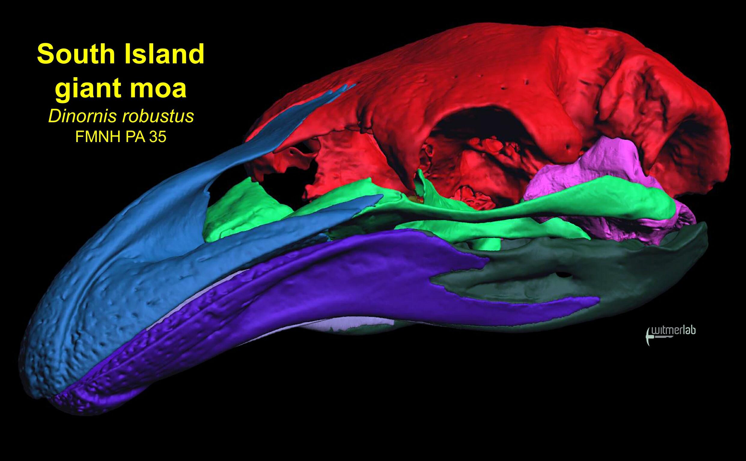
3D PDF of the skull of a moa (Dinornis
robustus: FMNH PA 35) with each
separable bony element as a separate colored object. In
this mature individual, many of the bones are fused.
•
Download a
41 MB 3D PDF LARGEST
•
Download a
28 MB 3D PDF LARGE
•
Download a
14 MB 3D PDF MEDIUM
•
Download a
7.7 MB 3D PDF SMALL
•
Download a
2 MB 3D PDF SMALLEST |
| |
|
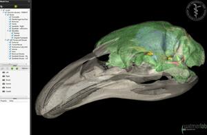 |
3D PDF of the skull of a moa (Dinornis
robustus: FMNH PA 35) with soft tissues such as the brain endocast,
inner ear labyrinth, and pneumatic sinuses in the
braincase and quadrate.
•
Download a
67 MB 3D PDF LARGEST
•
Download a
26 MB 3D PDF LARGE
•
Download a
13 MB 3D PDF MEDIUM
•
Download a
6.6 MB 3D PDF SMALL
•
Download a
4.2 MB 3D PDF SMALLEST |
| |
|
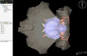 |
3D PDF of the braincase of a moa (Dinornis
robustus: FMNH PA 35) with soft tissues such as the
brain endocast, inner ear labyrinth, nerves, and blood
vessels.
•
Download a 30 MB 3D PDF LARGEST
•
Download a
24 MB 3D PDF LARGE
•
Download a
13 MB 3D PDF MEDIUM
•
Download a
7 MB 3D PDF SMALL
•
Download a
4.5 MB 3D PDF SMALLEST |
|
|
|
Labeled animation of skull, brain endocast, and inner ear. Animation of the skull of an adult
female of the extinct South Island giant moa (Dinornis
robustus, FMNH PA 35), labeled to show the endocast of the brain cavity, labyrinth of the inner ear,
confluent paranasal and paratympanic air sinuses, and other soft tissues.
It was collected in 1949 in the Pyramid Valley swamp near
Christchurch, New Zealand, for the Canterbury Museum. Work on this project was done by WitmerLab PhD student
Catherine Early. The bones of the braincase, mandible, and face
were CT scanned at OhioHealth O’Bleness Hospital, Athens, Ohio,
at a slice thickness of 300 µm, and the quadrate bone was
scanned at the OUµCT facility at a slice thickness of 45 µm. Segmentation of anatomical structures was done using Avizo; 3D PDFs were generated using Maya, Deep Exploration, and Adobe Acrobat; and movies were made using Avizo,
Maya, QuickTime, and Adobe Premiere.
•
Download a
62 MB QuickTime version (HD: 1920x1080)
•
Download a
33 MB QuickTime version (1280x720)
•
Download a
19 MB QuickTime version (853x480)
•
Download a
13 MB QuickTime version (640x360) |
|
|
|
|
Labeled skull animation.
Animation of the skull of an
adult female of the extinct
South Island giant moa (Dinornis
robustus, FMNH PA 35), labeled to show the
individual bones of the skull.
In this mature individual, many
of the bones are fused. It was
collected in 1949 in the Pyramid
Valley swamp near Christchurch,
New Zealand, for the Canterbury
Museum. Work on this project was done by WitmerLab PhD student
Catherine Early. The bones of
the braincase, mandible, and
face were CT scanned at
OhioHealth O’Bleness Hospital,
Athens, Ohio, at a slice
thickness of 300 µm, and the
quadrate bone was scanned at the
OUµCT facility at a slice
thickness of 45 µm. Segmentation of anatomical structures was done using Avizo; 3D PDFs were generated using Maya, Deep Exploration, and Adobe Acrobat; and movies were made using Avizo,
Maya, QuickTime, and Adobe Premiere.
•
Download a
21 MB QuickTime
version (HD: 1920x1080)
•
Download an
11 MB QuickTime
version (1280x720)
•
Download a
6 MB QuickTime
version (853x480)
•
Download
a 4.1 MB QuickTime
version (640x360) |
|
|
|
|
|
|
|