Common
Language Summary
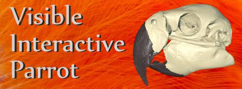
The Visible Interactive Parrot.
This page presents our work on the 3D anatomical
structure of the head and skull of the scarlet macaw, Ara
macao. Macaws, like all parrots, are unique among birds in
their intelligence and jaw anatomy (e.g., their remarkable
cranial kinesis), which makes them of particular interest to
members of WitmerLab. These resources are outgrowths of our more
technical work and are intended to serve as STEM educational
aids for K–12 and undergraduate students, as well as
researchers. Work on this project was done primarily by
WitmerLab PhD student JP Nassif. The featured specimen (OUVC
10633) is the dried skull of an adult scarlet macaw. The skull
was µCT-scanned with a voxel size of 90 µm (0.090 mm) at the
OUµCT facility. Segmentation of anatomical structures was done
using Avizo, 3D modeling was done using Maya, 3D PDFs were
generated using Deep Exploration and Adobe Acrobat, and movies
were made using QuickTime and Adobe Premiere. Other featured
specimens include a hyacinth macaw, and more content will
be added in the future. Funded by the National Science
Foundation.
Check out
our otherVisible Interactive Anatomy sites!
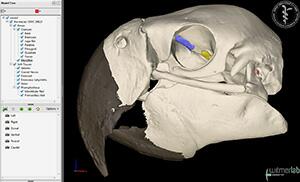
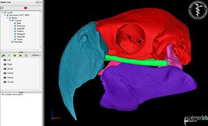
3D PDFs allow anyone with even the free Acrobat
Reader to interactively manipulate the 3D models that we
generate with powerful software like Avizo. The skull
and individual bones can be spun around, isolated, made
transparent, hidden, etc. The files can even be saved to
your local computer. We provide each 3D PDF in different resolutions and files sizes to match your
interest and the power of your computer.
View our mini-tutorial. NOTE: Bugs in many browsers prevent them from running
3D PDFs in a browser window, so please save it to your
system and then launch it.
Labeled animation of skull, brain endocast, and inner ear.
Animation of the skull of an
adult scarlet macaw (Ara
macao, OUVC 10633) labeled
to show the endocast of the
brain cavity, labyrinth of the
inner ear, and other soft
tissues. Work on this project
was done primarily by WitmerLab
PhD student James Nassif. The
skull was µCT-scanned with a
voxel size of 90 µm (0.090 mm)
at the OUµCT facility. Digital
extraction of anatomical
structures was done using Avizo;
3D modeling was done using Maya;
3D PDFs were generated using
Maya, Deep Exploration, and
Adobe Acrobat; and movies were
made using Avizo, Maya, and
Adobe Premiere.
•
Download a
30 MB MP4 movie (HD: 1920x1080)
•
Download a
14 MB MP4 movie (1280x720)
•
Download a
9 MB MP4 movie (853x480)
•
Download a
4 MB MP4 movie (640x360)
Labeled skull animation. Animation of the skull of an adult scarlet macaw (Ara macao, OUVC 10633) labeled to show the individual bones of the skull. In this mature individual, many of the bones are fused, and the individual components reflect functional units associated with the cranial kinesis mechanism. Work on this project was done primarily by WitmerLab PhD student James Nassif. The skull was µCT-scanned with a voxel size of 90 µm (0.090 mm) at the OUµCT facility. Digital extraction of anatomical structures was done using Avizo; 3D modeling was done using Maya; 3D PDFs were generated using Maya, Deep Exploration, and Adobe Acrobat; and movies were made using Avizo, Maya, and Adobe Premiere.
• Download a 19 MB MP4 movie (HD: 1920x1080)
• Download a 9 MB MP4 movie (1280x720)
• Download a 5 MB MP4 movie (853x480)
• Download a 2.5 MB mp4 MP4 movie (640x360)
Cranial kinesis in a hyacinth macaw. Demonstration of cranial kinesis in a hyacinth macaw, the largest flying parrot. WitmerLab doctoral student James Nassif dissected this specimen (OUVC 10883), and it was then skeletonized by our dermestid beetle colony. The video was made immediately after the skull came out of the disinfecting hydrogen-peroxide/ammonia bath, while the skull joints were still flexible. The amount of prokinesis is probably somewhat exaggerated compared to in life, but parrots indeed have extreme cranial kinesis.
• Download a 72 MB QuickTime movie (HD: 1920x1080)
Witmer is responsible for
the content of the website. Content provided here is for
educational and research purposes only, and may not be used for
any commercial purpose without the permission of
L. M. Witmer and other
relevant parties.