|
Lawrence M. Witmer,
PhD
Professor of Anatomy
Chang Professor of Paleontology
Dept. of Biomedical Sciences
Heritage
College of Osteopathic Medicine
Life Science Building, Rm 123
Ohio University
Athens, Ohio 45701 USA
Phone: 740 593 9489
Fax: 740 593 2400
Email:
witmerL@ohio.edu
|
|
|
|


 |
|
|
|
| |
|
Common
Language Summary
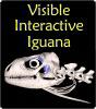
The Visible Interactive Iguana.
This page presents our work on the 3D anatomical
structure of the head and skull of the green iguana,
Iguana iguana. These resources are
outgrowths of our more technical work and are intended to serve
as STEM educational aids for K-12 and undergraduate students, as
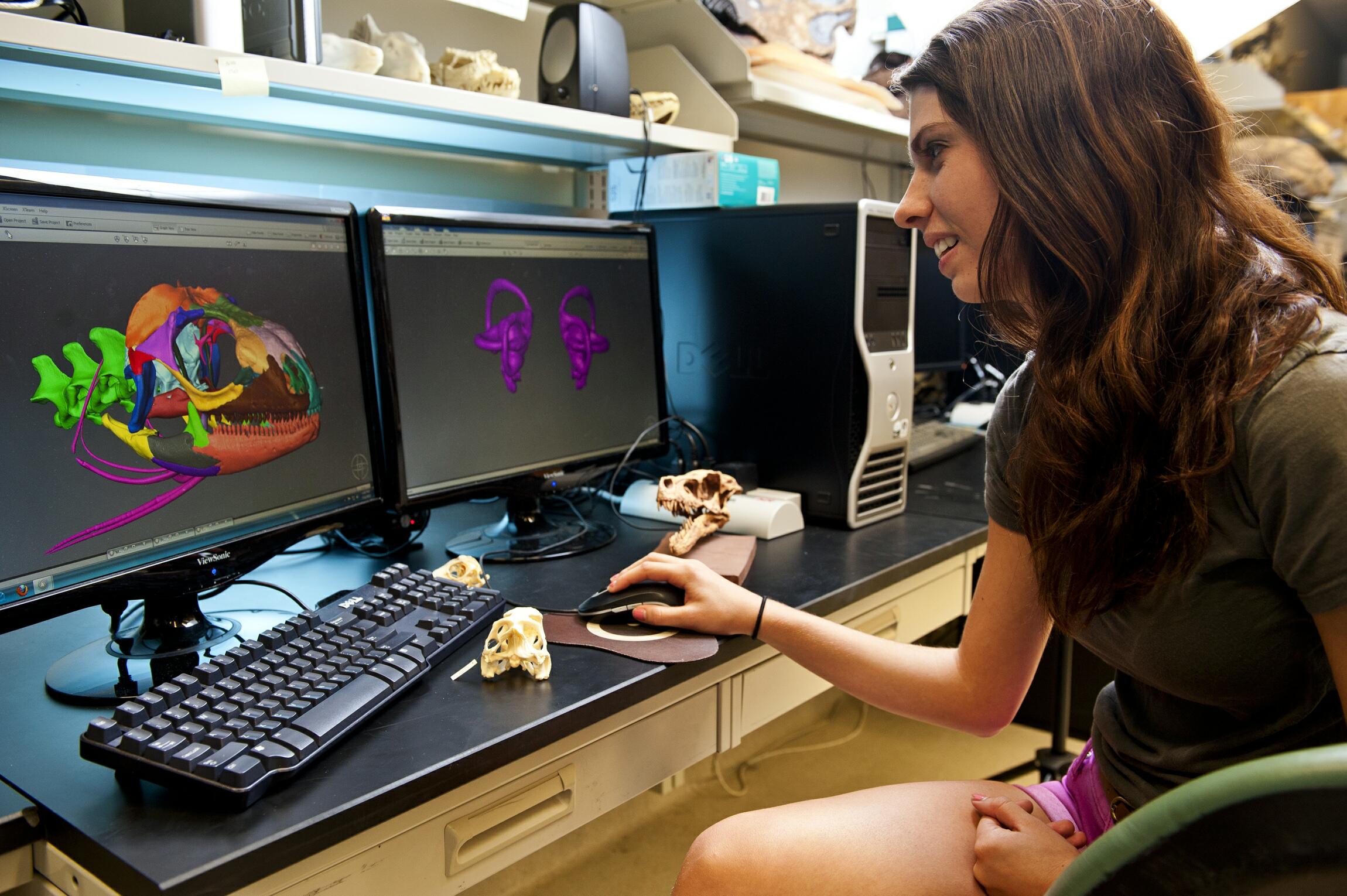
well as researchers. Our featured specimen is an adult green
iguana (OUVC 10677) collected legally and euthanized humanely in
Zoo
Miami and provided to us for research and
education. We scanned the head region on
the
OUµCT scanner in 2012 at a resolution of 90µm (0.090
mm). We
imported the scan data into
workstations running Avizo and digitally
extracted the bones and
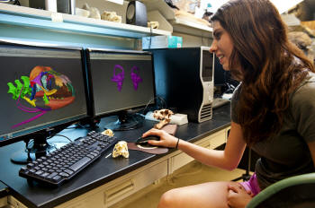
soft tissues. The work on this project was done
primarily by Alexandra Spaw Johns, an undergraduate student in
Ohio
University's Honors Tutorial College as part of her 2012
Summer Research Apprenticeship in WitmerLab. Lexie segmented all
the structures in Avizo, generated the 3D PDFs in Deep
Exploration and Adobe Acrobat, and made the movies in Adobe
Premiere. She was assisted especially by Jason Bourke, as well
as other WitmerLab members. More content will be added in the
future. |
|
|
|
|
 Download the µCT scan data
in DICOM format and 3D-printable STLs at MorphoSource.org
Download the µCT scan data
in DICOM format and 3D-printable STLs at MorphoSource.org
|
|
Some of the work
featured here has
been published:
•
Porter, W. R. and L. M. Witmer. 2015. Vascular patterns in
iguanas and other squamates: blood vessels and sites of thermal
exchange. PLOS ONE 10(10): e0139215.
doi:10.1371/journal.pone.0139215. 3D
PDF download
here. DICOM
data download for five OUVC specimens on
Dryad. |
|
Sketchfab
animations |
|
|
|
3D PDFs |
Videos |
3D PDFs allow anyone with even the free Acrobat
Reader to interactively manipulate the 3D models that we
generate with powerful software like Avizo. The skull
and individual bones can be spun around, isolated, made
transparent, hidden, etc. The files can even be saved to
your local computer. We provide each 3D PDF in different resolutions and files sizes to match your
interest and the power of your computer.
View our mini-tutorial.
NOTE: Bugs in many browsers prevent them from running
3D PDFs in a browser window, so please save it to your
system and then launch it.
|
|
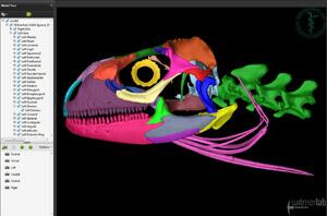 |
3D PDF of the skull of an adult
green iguana (Iguana iguana: OUVC
10677) with each
bone as a separate colored object. The right side and
left side can each be turned on and off or made
transparent. Unpaired bones (e.g., frontal) are assigned
to the right side.
•
Download a
13 MB 3D PDF LARGEST
•
Download a
7.2 MB 3D PDF LARGE
•
Download a
4.2 MB 3D PDF MEDIUM
•
Download a
2.5 MB 3D PDF SMALL |
| |
|
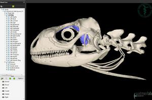 |
3D PDF of the skull of an adult
green iguana (Iguana iguana: OUVC
10677) with soft tissues such as the brain endocast,
inner ear labyrinth, blood vessels, and nerves. The
right side and left side can each be turned on and off
or made transparent. Unpaired bones (e.g., frontal) are
assigned to the right side.
•
Download a
14 MB 3D PDF LARGEST
•
Download a
7.7 MB 3D PDF LARGE
•
Download a
4.4 MB 3D PDF MEDIUM
•
Download a
2.6 MB 3D PDF SMALL |
| |
|
|
|
|
 |
Witmer is responsible for
the content of the website. Content provided here is for
educational and research purposes only, and may not be used for
any commercial purpose without the permission of
L. M. Witmer and other
relevant parties.
This project was funded by grants from the
National Science
Foundation. |
 |
|
|