Common
Language Summary
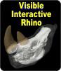
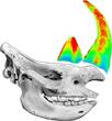
The Visible Interactive Rhino.
This page presents our work on the 3D anatomical
structure of the head and skull of white rhinoceros, Ceratotherium simum. These resources are
outgrowths of our work with the
Kariega Game
Reserve in South Africa. In March 2012, following the brutal
attack of three rhinos by poachers seeking horn, Dr. William
Fowlds, the wildlife veterinarian treating the rhinos, contacted
WitmerLab for insight into the anatomical structure of the horn,
skull, and nasal cavity of rhinos. The poachers had used
machetes to hack off the horns, leaving deep wounds in the face
and exposing the delicate mucous membranes of the paranasal air
sinuses and nasal cavity. WitmerLab provided Dr. Fowlds with the
anatomical information he requested, as well as generated
imagery that could be used in a more public context to help
highlight the extent of the injuries inflicted on rhinos by the
poachers. This web site seeks to share some of that imagery, as
well as to provide basic anatomical information that can serve
as STEM educational aids for K–12 and undergraduate students, as
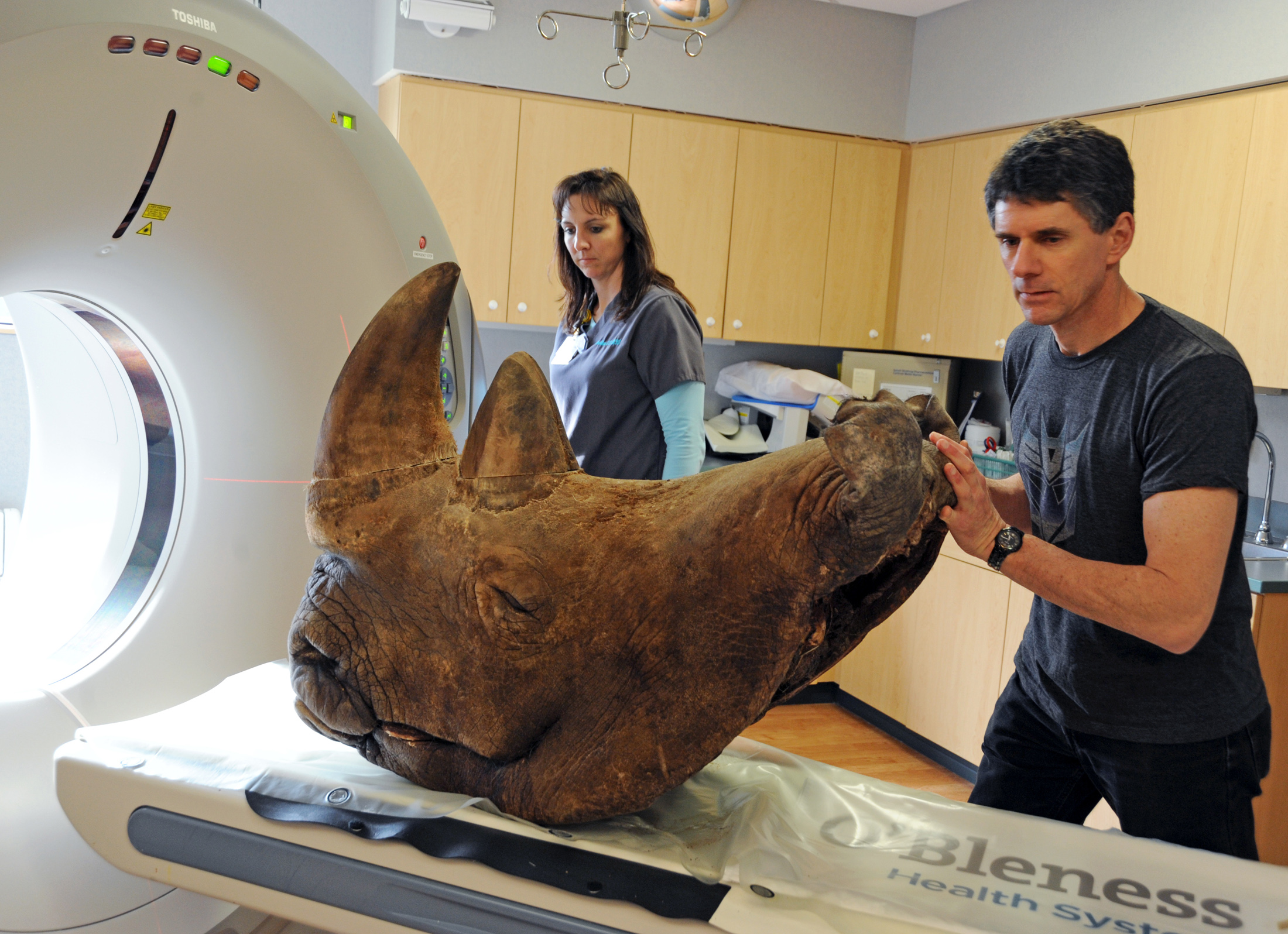
well as for researchers. Our primary specimen is the head of a
41-year-old male white rhino (named Kehtla) which was provided
to WitmerLab by the Phoenix Zoo in 2002. This specimen (OUVC
9754) contributed to a
published study on
rhino horn published by WitmerLab in 2006, the same study
that drew the attention of Dr. Fowlds. Kehtla's head was first
CT scanned with the help of Heather Rockhold, RT(CT), at
OhioHealth
O'Bleness Hospital in 2008 and then again in March 2012 so as
to get better data to help the Kariega rhinos. Chief WitmerLab
Research Associate Ryan Ridgely assembled the several sets of CT
scan data into a single 3D volume which is the basis for the
work here.
Check out
our other Visible Interactive Anatomy sites!
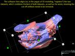
3D PDFs allow anyone with even the free Acrobat
Reader to interactively manipulate the 3D models that we
generate with powerful software like Avizo. The head, skull,
horns,
and soft tissues can be spun around, isolated, made
transparent, hidden, etc. The files can even be saved to
your local computer. We provide each 3D PDF in different resolutions and files sizes to match your
interest and the power of your computer.
View our mini-tutorial. NOTE: Bugs in many popular browsers prevent running
3D PDFs in a browser window, so you may need to save it to your
system and then launch it in Reader or Acrobat.
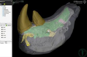
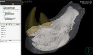
POACHED: 3D PDF of the
head of a 41-year-old male white rhinoceros (Ceratotherium
simum, OUVC 9754), replicating the effects of horn
poaching, with the skin, horns, skull, brain, eyeballs,
nasal cavity, and paranasal air sinuses as separate
objects. •
Download a
36 MB 3D PDF LARGEST •
Download a
17 MB 3D PDF LARGE •
Download a
9.5 MB 3D PDF MEDIUM •
Download a
5.0 MB 3D PDF SMALL
Videos from elsewhere
AMNH Science Bulletin.
Video characterizing WitmerLab's role in the wake of the
Kariega poaching incident, put together by Mindy
Weisberger and her Science Bulletin team at the American
Museum of Natural History.
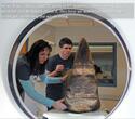
Athens Messenger.Video characterizing the partnership of O'Bleness
Memorial Hospital and WitmerLab in scanning Kehtla to
help the veterinary teams working at Kariega. The video
was put together by Athens Messenger reporter
Sara Brumfield.
Check out
Sara Brumfield's article in the
Athens Messenger.
Witmer, with the skilled
assistance of
Ryan Ridgely, is responsible for
the content of the website. Content provided here is for
educational and research purposes only, and may not be used for
any commercial purpose without the permission of
L. M. Witmer and other
relevant parties.