Common
Language Summary
The Visible Interactive Alligator Project at Ohio University and
the University of Missouri.
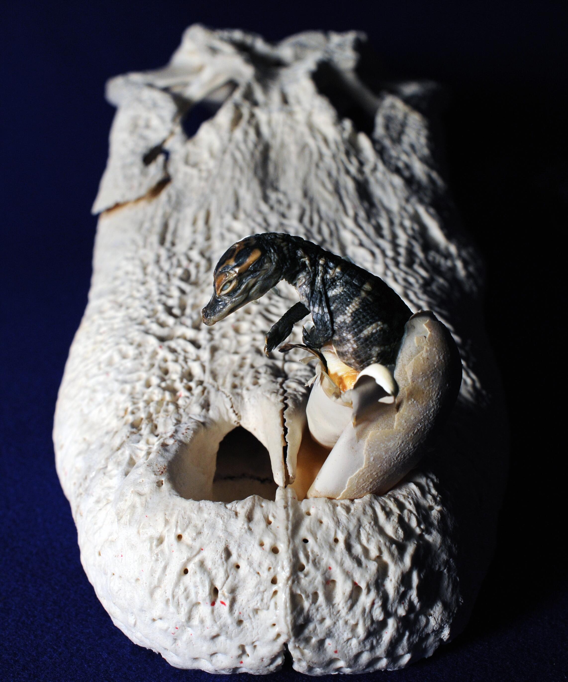
This page presents our work on the 3D anatomical
structure of the head and skull of American alligator,
Alligator mississippiensis. These resources are
outgrowths of our more technical work and are intended to serve
as STEM educational aids for K-12 and undergraduate students, as
well as researchers. Our specimens include a
"Day-0" hatchling, meaning it's from an animal that
was stillborn
on the day of hatching. We scanned the head region on
the
OUµCT scanner in 2009 at a resolution of 45µm (0.045
mm). We
imported the scan data into
workstations running Avizo and digitally
extracted the bones and
soft tissues.
See
the Pick-and-Scalpel blog post for details and to provide
feedback. Four
WitmerLab
doctoral students participated, principally
Dave Dufeau, who got the project started with the
braincase and soft tissues, and
Jason Bourke, who segmented the
rest of the bones
and made all the movies.
Ashley Morhardt and
Ruger Porter made the 3D PDFs. More recently, we scanned
a head of a subadult (OUVC 11415) at 50µm; this dataset was
visualized by Seishiro Tada of the University of Tokyo. We also include
some earlier WitmerLab work on an adult head. All of these
studies were funded by grants from the
National Science
Foundation (NSF). This work is done
in collaboration with the
Holliday Lab at the University of Missouri. They
have comparable content on their
3D Alligator site on an adult alligator.
Check out
our other Visible Interactive Anatomy sites!
3D PDFs allow anyone with even the free Acrobat
Reader to interactively manipulate the 3D models that we
generate with powerful software like Avizo. The skull
and individual bones can be spun around, isolated, made
transparent, hidden, etc. The files can even be saved to
your local computer. We provide each 3D PDF in three
different resolutions and files sizes to match your
interest and the power of your computer.
View our
mini-tutorial. NOTE: Bugs in many browsers prevent them from running
3D PDFs in a browser window, so please save it to your
system and then launch it.
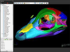
3D PDF of the skull of a day-0
hatchling Alligator
mississippiensis (OUVC
10606) with each
bone as a separate colored object. The right side and
left side can each be turned on and off or made
transparent. Unpaired bones (e.g., frontal) are assigned
to the right side.
•
Download a 22 MB 3D PDF LARGE
•
Download a 7.5 MB 3D PDF MEDIUM
•
Download a 3.5 MB 3D PDF SMALL
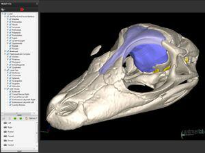
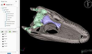
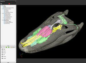
3D PDF of the skull of a day-0
hatchling Alligator
mississippiensis (OUVC
10606) with soft
tissues such as the brain endocast, inner ear labyrinth,
blood vessels, nerves, and, added in Sept 2015,
paratympanic air sinuses based on
Dufeau & Witmer (2015). Each bone is still a separate
object, but in this case, the bones are arranged in
anatomical groups (e.g., braincase), and individual
named bones (e.g., maxilla) can be turned on and off or
made transparent as left/right pairs.
•
Download a 25 MB 3D PDF LARGE
•
Download a 12 MB 3D PDF MEDIUM
•
Download a 6 MB 3D PDF SMALL
Witmer is responsible for
the content of the website. Content provided here is for
educational and research purposes only, and may not be used for
any commercial purpose without the permission of
L. M. Witmer and other
relevant parties.